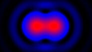
The light wavelength and numerical aperture determine the limit
In the simplest case, an optical microscope consists of one lens close to the specimen or sample (the objective) and one lens close to the eye (the eyepiece). The total microscope magnification is the product of the magnification factors of both lenses. For example, a 40x objective lens and a 10x eyepiece provide a total magnification of 400x.
However, it is not only magnification, but also resolution that indicates the performance capability of an optical microscope. Resolution is the ability to observe two closely adjacent points or dots as separate things. According to the Rayleigh criterion, the minimum distance between the two dots which allows them to be distinguished during imaging corresponds to approximately six tenths of the wavelength of the light [1].

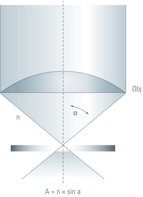
λ = wavelength of light
n = refractive index of the medium between the specimen or sample and objective
α = half the aperture angle of the objective
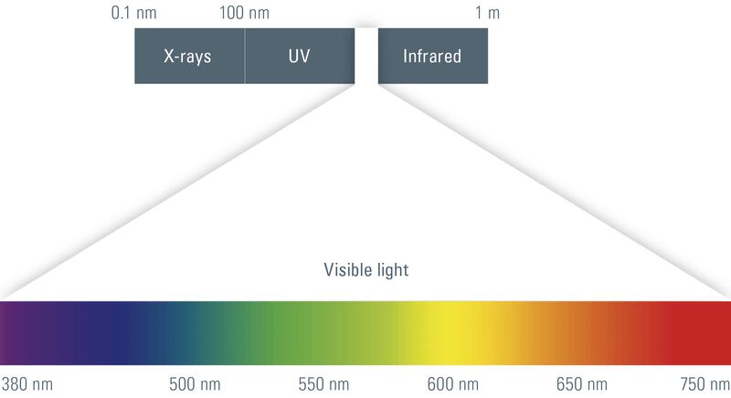
Therefore, with violet light the resolution limit is approximately d = 0.17 μm, with white light (average wavelength is 550 nm) d = 0.24 μm, and with red light d = 0.31 μm. UV objectives can attain a resolution of 0.15 μm. With the naked eye, most people are not able to differentiate structures smaller than 0.1 to 0.25 mm. When viewing images via the microscope eyepieces, then white light is always used. For UV, violet, or red light, then digital microscope cameras would be required.
The value n × sin α corresponds to the numerical aperture (NA) which is the measure of the light gathering capacity and the resolution of an objective lens. Because the aperture angle cannot exceed 90° and the refractive index is never less than 1 (nair = 1), the maximum NA is always around 1 for air. When immersion oil is used (n > 1), the maximum NA increases up to approximately 1.45 and the resolution along with it.
More magnification is not always better
The microscope’s resolution and magnification are always interdependent. An objective with low magnification has a small numerical aperture (NA) and, thus, a low resolution. A high-magnification objective has a large NA, typically 0.8 for a 40x objective which images in air. However, because the NA cannot be increased beyond a certain point, the useful magnification range of optical microscopes [2,3] is also limited. Depending on the wavelength of light used for illumination and the NA, the "useful" range of microscope magnification is shown in tables 1 and 2 below.
| Light Wavelength (λ, nm) | Useful Magnification Range (UMR) |
|---|---|
| 550 (white) | 500 × NA < UMR < 1,000 × NA |
| 400 (violet) | 700 × NA < UMR < 1,400 × NA |
| 340 (UV) | 800 × NA < UMR < 1,600 × NA |
Table 1: The useful magnification range (UMR) for an optical microscope depends on the wavelength of light used for illumination and the numerical aperture (NA) of the objective lens. N.B.: With white light, images can be viewed via the microscope eyepieces, but with UV and violet light, then a digital microscope camera must be used.
NA
NA | Light Wavelength (λ, nm) | ||
|---|---|---|---|
| 550 (white) | 400 (violet) | 340 (UV) |
| Useful Magnification Range (UMR) | ||
0.95 | 475x < UMR < 950x | 665x < UMR < 1,330x | 760x < UMR < 1,520x |
1 | 500x < UMR < 1,000x | 700x < UMR < 1,400x | 800x < UMR < 1,600x |
1.3 | 650x < UMR < 1,300x | 910x < UMR < 1,820x | 1,040x < UMR < 2,080x |
1.4 | 700x < UMR < 1,400x | 980x < UMR < 1,960x | 1,120x < UMR < 2,240x |
Table 2: The UMR (refer to table 1 above) for several higher NA values is shown here. N.B.: With white light, the microscope eyepieces can be used, but UV and violet light require a digital microscope camera.
Some optical microscopes, especially digital microscopes where the image is displayed on an electronic monitor, boast enormous magnification, but practically speaking, the limit for visible light (refer to figure 1) is just under 2,000x (for an NA of 1.4, refer to table 2) [2,3]. Any value beyond the useful range is called empty magnification, because specimen and sample structures appear larger, but no additional details are resolved.


