
Revealing Neuronal Migration’s Molecular Secrets
In this webinar, explore the molecular mechanisms of cortex development – from protein structure to neuronal migration.

17 October 2024 | 14:00 London, 15:00 Berlin, 17:00 Dubai
Different approaches can be used to investigate neuronal migration to their niche in the developing brain. In this webinar, experts from The University of Oxford present the microscopy tools and assays they use to elucidate the molecular mechanisms of neuronal migration to functional layers of the cortex during neurodevelopment. Understanding these processes will lead to a better understanding of healthy brain development and potentially better treatments for neurodevelopmental disorders.
Key webinar learnings:
- How different microscopy techniques are used to explore the role of receptors in guiding brain development.
- How cell biology assays such as stripe, cell aggregation and surface binding assays can help investigate the functions of receptors and their complexes in vitro.
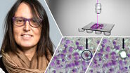
- How the THUNDER Imaging System within The University of Oxford’s Micron Bioimaging Facility supports this research.
Understanding molecular interactions during cortical development
The cortex is a many-layered and complex area of the brain. During neurodevelopment, the formation of a layered cortex requires the migration of neurons from where they arise, to their functional layer. This cortical migration requires precisely regulated interactions between cell adhesion receptors. These are proteins that interact with other molecules to guide neurons to their niche location.
In recent years, Professor Elena Seiradake’s team have functionally characterised several protein complexes that create a network of cell adhesion receptors. They use a combination of structural biology (macromolecular crystallography and cryo-EM) and cellular biology to create tools that enable them to study the function of each complex in vitro and in vivo.
In this webinar, members of Elena’s team will present case studies exploring the microscopy-based tools and assays they use to elucidate some of these complex interactions, and how these can contribute to wider understanding of the biology of neuron migration. These include surface binding assays, aggregation assays, and stripe assays, as well as sample preparation for cryo-electron tomography.
This webinar is being organized in collaboration with the Royal Microscopical Society (RMS).
Register for this webinar on the RMS website:
Related Articles
-

Probing Human Alzheimer's Cortical Section using Spatial Multiplexing
Alzheimer’s disease (AD) is the most common neurodegenerative disease and is characterized by the…
Sep 17, 2024Read article -
.")
Neuron Isolation in Spatial Context with Laser Microdissection (LMD)
After Alzheimer’s disease, Parkinson’s is the second most common progressive neurodegenerative…
Jun 24, 2024Read article -
How did Laser Microdissection enable Pioneering Neuroscience Research?
Dr. Marta Paterlini, a Senior Scientist at the Karolinska Institute, shares her experience of using…
Jun 24, 2024Read article
