Join us at FOM 2025
Learn how innovative new imaging systems from Leica Microsystems can help drive new discoveries in your research. Schedule your booth visit today!
- Discover the power of 3D high-plex imaging across scales : 15+ markers in one go!
- Uncover deeper cellular insights by combining THUNDER with spinning disk in the enhanced, game-changing, DMi8 inverted microscope
- Illuminate life in its entirety: exploring deep, long-term imaging with Viventis Deep
JOIN US!
FOM 2025
Leica Microsystems Booth #48
Exhibit Dates: April 13-16, 2025 | Taipei City, Taiwan

NEW PRODUCT PRESENTATIONS!
Overcoming the high multiplexing barrier: 3D Spatial Omics in one go
Sunday, April 13th, 2025 | 09:30 – 10:30 AM
Room 202
Speakers:
Dr. Taryn Guinan, Senior Application Specialist and Business Development Manager
In this presentation, we will elaborate on the experimental design and considerations for performing high multiplexing imaging experiments beyond 15+ fluorophores on a single sample. We will discuss appropriate fluorophore panels, details of sample preparation, and the challenges and tools required for imaging. Furthermore, Aivia Leica's cutting-edge image analysis software enables robust and efficient analysis of high multiplexing imaging data, providing researchers with powerful tools to extract meaningful insights from complex spatial biology experiments.
Live imaging of large multicellular systems with open top dual view light sheet microscopy
Sunday April 13th, 2025 | 4:00 - 5:00 PM
Monday April 14th, 2025 | 10:30 – 11:30 AM
Room 202
Speakers:
Dr. Taryn Guinan, Senior Application Specialist and Business Development Manager
Visualizing individual cells within large 3D multicellular systems, such as developing organisms or 3D cultures like spheroids and organoids, is challenging due to light scattering, which limits penetration. Here, we introduce Viventis Deep: an open-top dual-view and dual-illumination light-sheet microscope purposefully designed for live imaging of large specimens at single-cell resolution. The configuration of objectives together with a customizable multiwell mounting system combines for the first-time dual view light sheet imaging with multiposition imaging.
Sign up for a reminder
Gain deeper insights through advanced automation to streamline imaging workflows
Tuesday, April 15th, 2025 | 1:00 – 2:00 PM
Room 202
Speakers:
Dr. Adam Cliffe, Market Development Manager
In this talk, we will introduce the new, enhanced DMi8 inverted microscope and its expanded functionality and intelligent automation, which streamlines complex workflows. Additionally, high-speed confocal imaging with the new Spinning Disk Confocal Scanner, combined with technologies such as THUNDER and Aivia AI Image Analysis, offers deeper cellular insights from 3D and live samples. Don’t miss this exciting session to see how these fundamental improvements allow you to focus more on producing quality, publishable data.
Sign up for a reminder
Join us for poster presentations!
Advances in Label-Free Chemical Imaging with Coherent Raman Scattering Microscopy
Monday April 14th, 2025 | 5:50 pm – 7:20 PM
Abstract Number: P1-A/13
Session Title: Vibration & Non-linear
Authors:
Dr. Adam Cliffe, Taryn Guinan and Volker Schweikhard
Sign up for a reminder
Open top multi sample dual view light sheet microscope for live imaging of large multicellular systems
Tuesday April 15th, 2025 | 5:50 pm – 7:20 PM
Abstract Number: P2-F/7
Session Title: Light Sheet & Correlative
Authors:
Taryn Guinan, Adam Cliffe, Franziska Moos, Gustavo de Medeiros, Prisca Liberali, Simon Suppinger, Petr Strnad, Andrea Boni, Judith Reddington
Can you Beat AI?
Test your cell counting skills against Mateo FL’s AI-powered cell counter! If your estimate is within 10% of the actual count, you'll earn a "Cell Counting Champ" sticker and enjoy unlimited bragging rights among your peers.
Visit booth 48 to play!
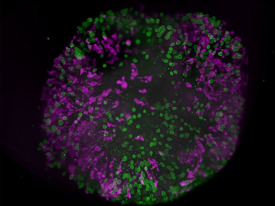
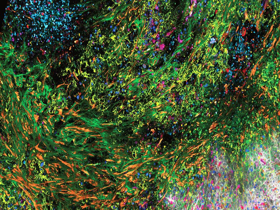
Next Gen STELLARIS - Discover the power of 3D high-plex imaging across scales
The next generation STELLARIS confocal platform with SpectraPlex allows you to significantly expand your spatial discoveries across scales! Discover new cell types, identify cell states, and map functional relationships in a spatial context to understand disease progression and identify potential therapeutic targets.
With a fully integrated 3D spatial imaging and analysis workflow (in combination with Aivia), it is no longer necessary to have a dedicated additional instrument to acquire multiplexed images that require cumbersome steps to analyze information from high-multiplexed samples.
The next generation STELLARIS brings you
- Access 3D spatial information with 15+ markers in one go: Expand captured information across scales
- Design your experiment in advance: Explore and optimize panel options with the integrated functionalities
- Manage data with minimal human interaction: Get intelligent guidance for setting up acquisition and performing the experimental controls
Can’t wait to learn more? Sign up for a reminder to our product presentation or sign up for a demo!
Enhanced DMi8 Inverted Microscope
Advanced Imaging. Simplified.
Streamline your complex microscopy workflows and uncover deeper cellular insights with the enhanced DMi8 inverted microscope. Reliably generate high quality data with a system that you can tailor to your research requirements and budget.
- Advanced automation to efficiently perform complex workflows
Focus on your cell biology research, not the system. Gaining deeper insights becomes simple with intelligent automation features that allow you to efficiently set up your experiments, including game-changing patented adaptive immersion technology, automated sample finder and 22mm FOV.
- Trust your results with higher quality image data and analysis
Generate better quality data every time regardless of your experience level. Extract the most insights from your 3D samples with our advanced technology suite including THUNDER, Spinning Disc Confocal Scanner, SmartCORR, TIRF and Aivia.
- Flexible tailored solutions
Future-proof your experiments! The DMi8's modular structure allows you to tailor your system to fit your current needs and budget. Then add features such as THUNDER, climate control, TIRF, spinning disk modules and more as your research evolves.
Viventis Deep - Revealing life in its full context
The Viventis Deep light sheet fluorescence microscope combines multi-view and multi-position light-sheet imaging to illuminate life in its entirety. Begin your journey to discover deep, long-term imaging that reveals the intricate details and dynamics of biological systems.
- Explore life in depth: expand the spatio-temporal understanding of your sample to its full-depth
- Explore multiple living samples in parallel: the open-top configuration enables higher throughput and multi-position capabilities
- Explore life events with long-term imaging: the gentle light sheet provides high image quality while preserving sample viability.