Julia Roberti , Dr

Julia Roberti

Julia studierte Chemie an der Universität von Buenos Aires, wo sie sich mit Photochemie und analytischer Chemie beschäftigte. Sie konzentrierte sich auf die Charakterisierung der Lumineszenz von Lanthanidkomplexen und auf die Entwicklung von Bor-Quantifizierungsmethoden für die Bor-Neutroneneinfangtherapie (BNCT) von Krebs. Nach ihrem Abschluss ging sie nach Göttingen, um am Max-Planck-Institut für Biologische Chemie zu promovieren. Sie entwickelte In-vitro- und In-situ-Fluoreszenzmarkierungsstrategien, um die Oligomerisierungs- und Aggregationsmechanismen des mit der Parkinsonschen Krankheit assoziierten Proteins Alpha-Synuclein aufzuklären. Im Jahr 2012 kam sie als Humboldt-Postdoc-Stipendiatin ans EMBL und setzte fortschrittliche konfokale Mikroskopie und Nanoskopie ein, um die Chromatinverdichtung am Übergang von der Interphase zur Mitose zu untersuchen. Seit 2017 ist sie bei Leica Microsystems als Produktmanagerin für fortschrittliche konfokale Bildgebung tätig.
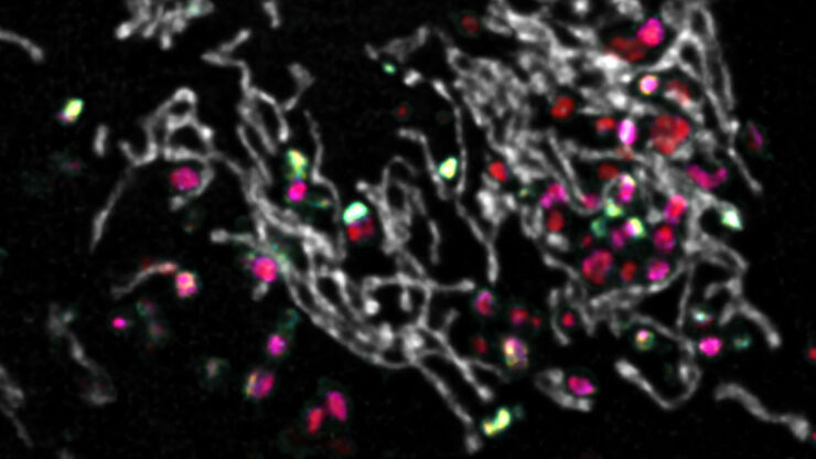
Insights into Vesicle Trafficking

STELLARIS provides integral access to complementary layers of information for dynamic, structural, and mechanistic insights into vesicle trafficking.
Spectral separation of 11 fluorophores coupled to polystyrene beads on a STELLARIS confocal system.

Multiplexing through Spectral Separation of 11 Colors

Fluorescence microscopy is a fundamental tool for life science research that has evolved and matured together with the development of multicolor labeling strategies in cells tissues and model…

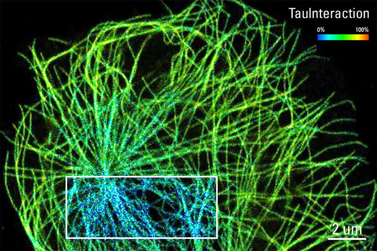
TauInteraction – Studying Molecular Interactions with TauSense

Fluorescence microscopy constitutes one of the pillars in life sciences and is a tool commonly used to unveil cellular structure and function. A key advantage of fluorescence microscopy resides in the…

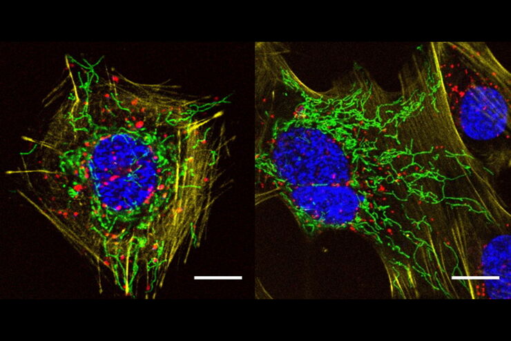
A Versatile Palette of Fluorescent Probes

Researchers at the Max Planck Institute for Medical Research in Heidelberg have developed a general strategy to synthesize live-cell compatible fluorogenic probes, and the result are the new MaP (Max…

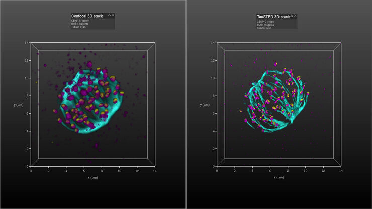
Kinetochore Assembly during Mitosis with TauSTED on 3D

Three-dimensional organization of the mitotic spindle together with the distribution of CENP-C and BUB1 based on TauSTED with multiple STED lines (592, 660 and 775 nm) can provide insights…
Scroll to top