Kontaktieren Sie uns

Leica Microsystems

Leica Microsystems

Leica Microsystems ist ein weltweit führender Hersteller von Mikroskopen und wissenschaftlichen Instrumenten. Im 19. Jahrhundert als Familienunternehmen gegründet, war die Geschichte des Unternehmens auf dem Weg zum Weltkonzern von beispielloser Innovation geprägt.

Die traditionell enge Zusammenarbeit mit der Wissenschaft ist der Schlüssel zur Innovationstradition von Leica Microsystems, die auf die Ideen der Anwender zurückgreift und auf deren Bedürfnisse zugeschnittene Lösungen schafft. Auf globaler Ebene ist Leica Microsystems in drei Divisionen gegliedert, die alle zu den führenden Anbietern in ihrem jeweiligen Bereich gehören: Life Science, Industrie und Medizin.

Das Unternehmen ist in über 100 Ländern mit 6 Produktionsstandorten in 5 Ländern, Vertriebs- und Serviceorganisationen in 20 Ländern und einem internationalen Händlernetz vertreten. Der Hauptsitz des Unternehmens liegt in Wetzlar, Deutschland.

https://www.leica-microsystems.com/de

Brief Introduction to Coating Technology for Electron Microscopy

Coating of samples is required in the field of electron microscopy to enable or improve the imaging of samples. Creating a conductive layer of metal on the sample inhibits charging, reduces thermal…

Carbon Thickness Evaluation in Electron Microscopy

The coating layers applied and used for electron microscopy imaging are commonly controlled and measured by quartz crystals. These crystals oscillate with a certain frequency (around 6 megahertz when…

Spectral Detection – How to Define the Spectral Bands that Collect Probe-specific Emission

To specifically collect emission from multiple probes, the light is first separated spatially and then passes through a device that defines a spectral band. Classically, this is a common glass-based…
Structural details of the C. elegans, head in cross-section. Courtesy of Müller-Reichert T, MPI-CBG, Dresden, Germany, and McDonald K, University of California, Berkeley, USA.

Brief Introduction to High-Pressure Freezing

Water is the most abundant cellular constituent and therefore important for preserving cellular ultra-structure. Currently the only way to fix cellular constituents without introducing significant…

Step by Step Guide for FRAP Experiments

Fluorescence Recovery After Photobleaching (FRAP) has been considered the most widely applied method for observing translational diffusion processes of macromolecules. The resulting information can be…
Forensics microscopy

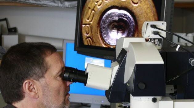
Every Clue Counts – Forensics Inconceivable Without Microscopy

There is no crime without clues. They may be obvious, like a cartridge case at the scene of the crime or clear signs of crowbar damage on a door. But sometimes, clues are microscopically small.…

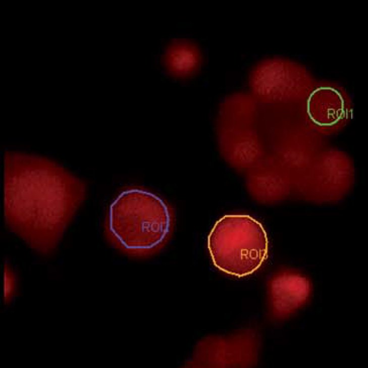
Widefield Calcium Imaging with Calcium Indicator Fura2

In eukaryotic cells Ca2+ is one of the most widespread second messengers used in signal transduction pathways. Intracellular levels of Ca2+ are usually kept low, as Ca2+ often forms insoluble…
Blood cells. Critical Point Drying.

Brief Introduction to Critical Point Drying

One of the uses of the Scanning Electron Microscope (SEM) is in the study of surface morphology in biological applications which requires the preservation of the surface details of a specimen. Samples…
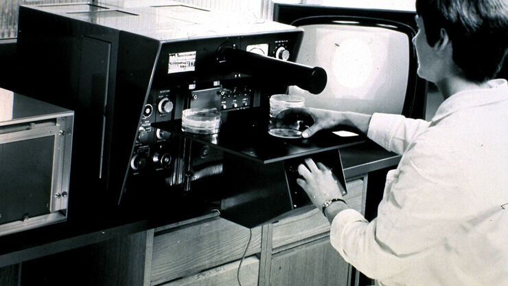
QTM B, 1963, the first commercial automated image analysis system for microscope images, based on a TV camera and developed by Metals Research in Cambridge, England.

50 Years of Image Analysis

Modern image analysis systems perform highly sophisticated image processing functions on images from an automated microscope and digital camera. 50 years ago, the first image analysis system was…
Center a fluorescence bulb.

Video Tutorial: How to Align the Bulb of a Fluorescence Lamp Housing

The traditional light source for fluorescence excitation is a fluorescence lamp housing with mercury burner. A prerequisite for achieving bright and homogeneous excitation is the correct centering and…
Exchange a fluorescence bulb.

Video Tutorial: How to Change the Bulb of a Fluorescence Lamp Housing

When applying fluorescence microscopy in biological applications, a lamp housing with mercury burner is the most common light source. This video tutorial shows how to change the bulb of a traditional…
Sub-Femtolitre volume_Fluorescence correlation spectroscopy (FCS)

Fluorescence Correlation Spectroscopy (FCS)

Fluorescence correlation spectroscopy ( FCS ) measures fluctuations of fluorescence intensity in a sub-femtolitre volume to detect such parameters as the diffusion time, number of molecules or dark…
White Light Laser: Lambda square scan

White Light Laser

The perfect light source for confocal microscopes in biomedical applications has sufficient intensity, tunable color and is pulsed for use in lifetime fluorescence. Furthermore, it should offer means…

Controlling the TIRF Penetration Depth is Mandatory for Reproducible Results

The main feature of total internal reflection fluorescence (TIRF) microscopy is the employment of an evanescent wave for the excitation of fluorophores instead of using direct light. A property of the…
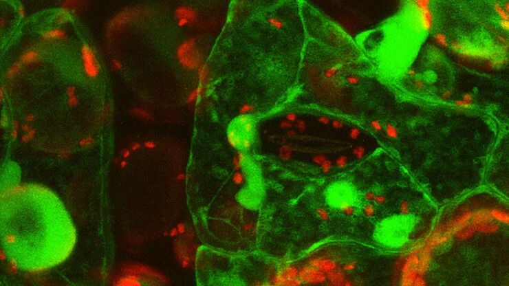
Primary leaves of cowpea (Vigna unguiculata "California Blackeye") inoculated with cowpea mosaic virus (CPMV) containing the GFP-gene inserted between the movement protein (MP) and the capsid proteins (CPs) in the viral RNA 2

Introduction to Live-Cell Imaging

The understanding of complex and fast cellular dynamics is an important step to get insight into biological processes. Therefore, today’s life science research more and more demands studying…
TIRF Image of Tubulin, YFP, penetration depth: 120 mm

Applications of TIRF Microscopy in Life Science Research

The special feature of TIRF microscopy is the employment of an evanescent field for fluorophore excitation. Unlike standard widefield fluorescence illumination procedures with arc lamps, LEDs or…
Jellyfish Aequorea Victoria

Fluorescent Proteins - From the Beginnings to the Nobel Prize

Fluorescent proteins are the fundament of recent fluorescence microscopy and its modern applications. Their discovery and consequent development was one of the most exciting innovations for life…
Snapshot from a time lapse of a calcium imaging experiment using the ratiometric calcium indicator Fura-2.

Ratiometric Imaging

Many fundamental functions of a cell strongly depend on delicate, but nevertheless dynamic balances of ions (e.g. calcium, magnesium), voltage potentials and pH between the cell’s cytosol and the…
Scroll to top