Leica Microsystems

Leica Microsystems

Leica Microsystems ist ein weltweit führender Hersteller von Mikroskopen und wissenschaftlichen Instrumenten. Im 19. Jahrhundert als Familienunternehmen gegründet, war die Geschichte des Unternehmens auf dem Weg zum Weltkonzern von beispielloser Innovation geprägt.

Die traditionell enge Zusammenarbeit mit der Wissenschaft ist der Schlüssel zur Innovationstradition von Leica Microsystems, die auf die Ideen der Anwender zurückgreift und auf deren Bedürfnisse zugeschnittene Lösungen schafft. Auf globaler Ebene ist Leica Microsystems in drei Divisionen gegliedert, die alle zu den führenden Anbietern in ihrem jeweiligen Bereich gehören: Life Science, Industrie und Medizin.

Das Unternehmen ist in über 100 Ländern mit 6 Produktionsstandorten in 5 Ländern, Vertriebs- und Serviceorganisationen in 20 Ländern und einem internationalen Händlernetz vertreten. Der Hauptsitz des Unternehmens liegt in Wetzlar, Deutschland.

https://www.leica-microsystems.com/de

Optical microscope image of salt contamination on an aluminum/silicon (Al/Si) surface. Credit: Gerweck GmbH, Germany.

Microscopic Defects in Electroplating

This free on-demand webinar shows how to identify quickly root causes of defects during electroplating of components with optical microscopy and LIBS (laser spectroscopy).

Insights into Vesicle Trafficking

STELLARIS provides integral access to complementary layers of information for dynamic, structural, and mechanistic insights into vesicle trafficking.

Visualizing Protein-Protein Interactions by Non-Fitting and Easy FRET-FLIM Approaches

The Webinar with Dr. Sergi Padilla-Parra is about visualizing protein-protein interaction. He gives insight into non-fitting and easy FRET-FLIM approaches.
Living HeLa cells stained with WGA-488 (yellow), SPY-Actin (cyan), and SiR-Tubulin (magenta). Instant Computational Clearing (ICC) was applied.

How to Perform Dynamic Multicolor Time-Lapse Imaging

Live-cell imaging sheds light on diverse cellular events. As many of these events have fast dynamics, the microscope imaging system must be fast enough to record every detail. One major advantage of…
Spectral separation of 11 fluorophores coupled to polystyrene beads on a STELLARIS confocal system.

Multiplexing through Spectral Separation of 11 Colors

Fluorescence microscopy is a fundamental tool for life science research that has evolved and matured together with the development of multicolor labeling strategies in cells tissues and model…

3 Factors Determine the Damage Potential of Particles

This article discusses the 3 factors for determining the potential of a particle to cause damage to parts and components in the automotive and electronic industry. These factors include the…
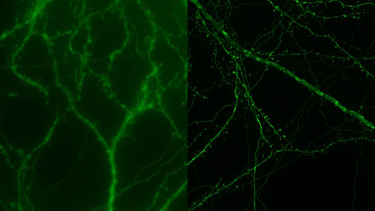
Raw widefield and THUNDER image of transversal mouse adult fiber lens section. Courtesy N. Houssin, Plagemen lab, Ohio State University, Columbus, USA.

Studying Ocular Birth Defects

This article discusses how lens formation and ocular birth defects can be studied with sharp widefield microscopy images which are acquired rapidly. The mouse ocular lens is used as a model to study…
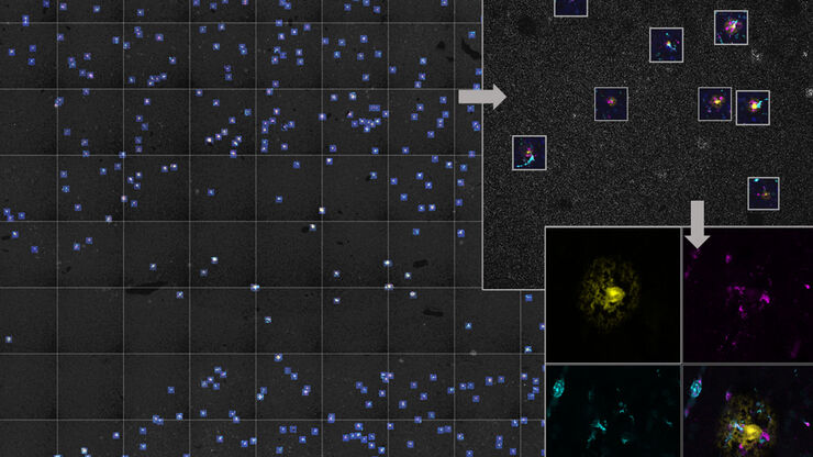
Two-color caspase assay with tile scan. U2OS cells were treated with the nuclear marker DRAQ5 (magenta) and CellEvent™ (yellow).

Following Multiple Events during Staurosporine Apoptosis

In this video on demand, we show how adding additional markers to an apoptosis kit can markedly increase the amount of information a researcher can obtain from the same experiment. The simultaneous…
Image of murine-brain tissue showing a region removed with UV laser microdissection.

RNA Quality after Different Tissue Sample Preparation

The influence of sample preparation and ultraviolet (UV) laser microdissection (UV LMD) on the quality of RNA from murine-brain tissue cryo-sections is described in this article. To obtain good…
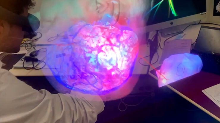
Analyse eines Aneurysmas mit Virtual Reality. Bild mit freundlicher Genehmigung von Prof. Raphael Guzman.

Digitalisierung in der neurochirurgischen Planung und bei Eingriffen

Erfahren Sie mehr über Augmented Reality, Virtual Reality und Mixed Reality in der Neurochirurgie und wie sie dazu beitragen können, Herausforderungen zu meistern.
The various solutions from Leica Microsystems for cleanliness analysis.

Factors to Consider for a Cleanliness Analysis Solution

Choosing the right cleanliness analysis solution is important for optimal quality control. This article discusses the important factors that should be taken into account to find the solution that best…
Projection of a confocal z-stack. Sum159 cells, human breast cancer cells kindly provided by Ievgeniia Zagoriy, Mahamid Group, EMBL Heidelberg, Germany. Blue–Hoechst - indicates nuclei, Green–MitoTracker mitochondria, and red–Bodipy - lipid droplets

New Imaging Tools for Cryo-Light Microscopy

New cryo-light microscopy techniques like LIGHTNING and TauSense fluorescence lifetime-based tools reveal structures for cryo-electron microscopy.
Transverse histological cut of a rabbit tongue. 50 Mpixels images (2326 µm x 1739 µm) in 14 x 18 tiles. Lifetime gives an additional contrast that allows to differentiate different structures in histological stainings.

Leitfaden zur Fluoreszenzlebensdauer-Imaging-Mikroskopie (FLIM)

Die Fluoreszenzlebensdauer ist ein Maß dafür, wie lange ein Fluorophor im Durchschnitt in seinem angeregten Zustand verbleibt, bevor er durch Aussendung eines Fluoreszenzphotons in den Grundzustand…
Staining is crucial to revealing details in the specimens.

H&E Staining in Microscopy

If we consider the role of microscopy in pathologists’ daily routines, we often think of the diagnosis. While microscopes indeed play a crucial role at this stage of the pathology lab workflow, they…
Untreated Hela Kyoto cells stained to show the nucleus (Hoechst, blue), the cis-golgi matrix protein GM130 (AF488, green), and the trans-golgi network membrane protein TGN46 (AF647, red).

Golgi Organizational Changes in Response to Cell Stress

In this video on demand, our special guest George Galea from EMBL Heidelberg will look at HeLa Kyoto cells treated with various chemotherapeutic agents to investigate their effect on the Golgi complex…
Type of contamination: spores

Cleanliness Analysis for Particulate Contamination

Devices, products, and their components fabricated in many industries can be quite sensitive to contamination and, as a result, have stringent requirements for technical cleanliness. Measurement…
Partikel und Fasern auf einem Filter, die gezählt und auf Sauberkeit analysiert werden.

Effiziente Partikelzählung und -analyse

Dieser Bericht befasst sich mit der Partikelzählung und -analyse unter Verwendung der optischen Mikroskopie bei der technischen Sauberkeitsanalyse von Teilen und Komponenten. Die Partikelzählung und…
Protist Paramecium (Paramecium tetraurelia) stained to show the nucleus

3D Spatial Analysis Using Mica's AI-Enabled Microscopy Software

This video offers practical advice on the extraction of publication grade insights from microscopy images. Our special guest Luciano Lucas (Leica Microsystems) will illustrate how Mica’s AI-enabled…
A stack of lithium-ion batteries

Quality Control Under the Microscope

Fast-rising demand for electric vehicles is one of the market’s main drivers, but there are other hotspots of growth, including the rise in renewable energy installations, such as photovoltaic panels,…
Pancreatic ductal adenocarcinoma tissue section imaged with Cell DIVE

Multiplexed Imaging Types, Benefits and Applications

Multiplexed imaging is an emerging and exciting way to extract information from human tissue samples by visualizing many more biomarkers than traditional microscopy. By observing many biomarkers…
3D Reconstruction of brain slide image_Mica

3D Tissue Imaging: From Fast Overview To High Resolution With One Click

3D Tissue imaging is a widespread discipline in the life sciences. Researchers use it to reveal detailed information of tissue composition and integrity, to make conclusions from experimental…
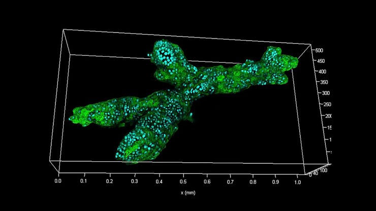
MDCK cysts on day 9

How To Perform Fast & Stable Multicolor Live-Cell Imaging

With the help of live-cell imaging researchers gain insights into dynamic processes of living cells up to whole organisms. This includes intracellular as well as intercellular activities. Protein or…
Image of a single slice taken from a zebrafish heart showing the ventricle with an injury in the lower area. Nuclei of all cells are indicated with blue, nuclei of the cardiomyocyte heart muscle cells with green, and the proliferating cells with red. Courtesy of Laura Peces-Barba Castaño, Max Planck Institute for Heart and Lung Research, Germany.

Imaging of Cardiac Tissue Regeneration in Zebrafish

Learn how to image cardiac tissue regeneration in zebrafish focusing on cell proliferation and response during recovery with Laura Peces-Barba Castaño from the Max Planck Institute.
When particulate contamination is present in lubricating fluids or oils, it can cause damage to parts or components leading to malfunctions.

Hydraulics in the Automotive and Aerospace Industries

Cleanliness standards relating to lubricants, hydraulic fluids, and oils, e.g., ISO 4406 and DIN 51455 are discussed in this article. Cleanliness plays a central role in the automotive and…

TauInteraction – Studying Molecular Interactions with TauSense

Fluorescence microscopy constitutes one of the pillars in life sciences and is a tool commonly used to unveil cellular structure and function. A key advantage of fluorescence microscopy resides in the…
Images of smooth muscle cells during wound healing. Courtesy L.S. Shankman, Ph.D., University of Virginia.

Studying Wound Healing of Smooth Muscle Cells

This article discusses how wound healing of cultured smooth muscle cells (SMCs) in multiwell plates can be reliably studied over time with less effort using a specially configured Leica inverted…
Partikel, die bei der Sauberkeitsanalyse von Teilen und Komponenten gefunden werden konnten.

Technische Sauberkeit von Automobilkomponenten und -teilen

In diesem Artikel werden die ISO-Norm 16232 und die VDA 19-Richtlinien erläutert und die Verfahren zur Partikelanalyse kurz zusammengefasst. Diese liefern wichtige Kriterien für die Sauberkeit von…

How Does The Cytoskeleton Transport Molecules?

VIDEO ON DEMAND - See how 3D cysts derived from MDCK cells help scientists understand how proteins are transported and recycled in tissues and the role of the cytoskeleton in this transport.
Developing zebrafish (Danio rerio) embryo, from sphere stage to somite stages.

Studying Early Phase Development of Zebrafish Embryos

This video on demand focuses on combining widefield and confocal imaging to study the early-stage development of zebrafish embryos (Danio rerio), from oocyte to multicellular stage.
Two-color caspase assay with tile scan

Multi-Color Caspase 3/7 Assays with Mica

Caspases are involved in apoptosis and can be utilized to determine if cells are undergoing this programmed cell death pathway in so-called caspase assays. These assays can be run by e.g. flow…
Scroll to top