Science Lab
Science Lab
Das Wissensportal von Leica Microsystems bietet Ihnen Wissens- und Lehrmaterial zu den Themen der Mikroskopie. Die Inhalte sind so konzipiert, dass sie Einsteiger, erfahrene Praktiker und Wissenschaftler gleichermaßen bei ihrem alltäglichen Vorgehen und Experimenten unterstützen. Entdecken Sie interaktive Tutorials und Anwendungsberichte, erfahren Sie mehr über die Grundlagen der Mikroskopie und High-End-Technologien - werden Sie Teil der Science Lab Community und teilen Sie Ihr Wissen!
Filter articles
Tags
Berichtstyp
Produkte
Loading...

Hochwertige EBSD-Probenvorbereitung
Es wird eine zuverlässige und effiziente EBSD-Probenvorbereitung von „gemischten“ kristallografischen Materialien mit Ionenbreitstrahlfräsen beschrieben. Das beschriebene Verfahren erzeugt…
Loading...

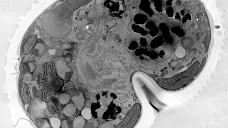
Wie die Analyse von Meeresmikroorganismen durch Hochdruckgefrieren verbessert werden kann
Die ultrastrukturelle Analyse von Umweltproben, hier Dinoflagellaten, bleibt heutzutage eine Herausforderung. Hier zeigen wir, dass die Durchführung von Hochdruckgefrieren (HPF) vor Ort die…
Loading...

Use of AR Fluorescence in Neurovascular Surgery
Learn about the use of GLOW800 Augmented Reality in neurovascular surgery through clinical cases and videos, including aneurysm and tumor resection cases.
Loading...

Life-Science-Forschung: Welche Mikroskopkamera ist die richtige für Sie?
Wie Sie entscheiden, welche Kamera für Ihre Life-Science-Mikroskopie-Experimente die Richtige ist. Welche Kamera von Leica Microsystems ist für Sie am besten geeignet?
Loading...

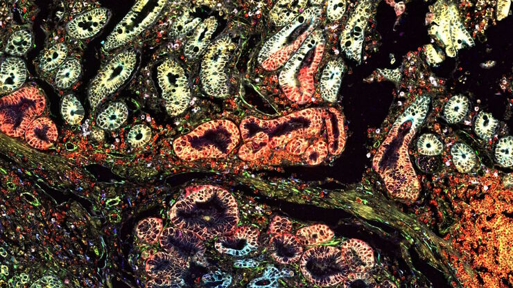
Multiplexing with Luke Gammon: Advance your Spatial Biology Research
Learn how multiplexing imaging and spatial biology can help researchers better understand complex biological systems. In this interview, Dr. Gammon and Dr. Pointu of Leica Microsystems discuss pain…
Loading...

Räumliche Biologie: Erwägung neuer Wege
Räumliche Biologie: Forschung zu Anordnung und Interaktion von Molekülen, Zellen und Geweben in ihrem nativen räumlichen Kontext
Loading...

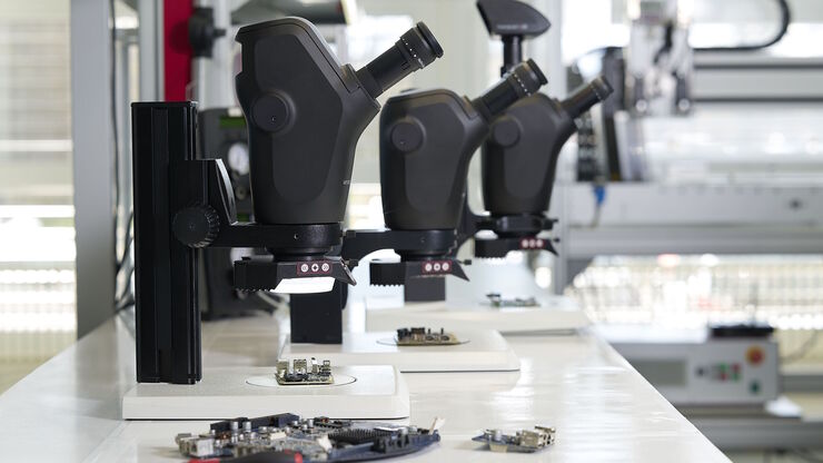
Wichtige Faktoren, die Sie bei der Auswahl eines Stereomikroskops berücksichtigen sollten
Stereomikroskope zeichnen sich durch ihre Fähigkeit aus, einen 3D-Eindruck der Probe zu erzeugen. Daher eignen sie sich besonders gut für Inspektion und Nacharbeit, Qualitätskontrolle, Forschung und…
Loading...

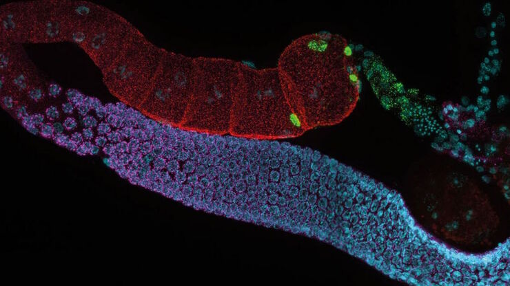
Imaging Organoid Models to Investigate Brain Health
Imaging human brain organoid models to study the phenotypes of specialized brain cells called microglia, and the potential applications of these organoid models in health and disease.
Loading...

Schnelle und zuverlässige Untersuchung von Leiterplatten und Leiterplattenbaugruppen mittels Digitalmikroskopie
Digitalmikroskope bieten Anwendern eine bequeme und schnelle Möglichkeit zur Erfassung hochwertiger, zuverlässiger Bilddaten und zur schnellen Inspektion und Analyse von Leiterplatten (PCBs) und…
