
Science Lab
Science Lab
Das Wissensportal von Leica Microsystems bietet Ihnen Wissens- und Lehrmaterial zu den Themen der Mikroskopie. Die Inhalte sind so konzipiert, dass sie Einsteiger, erfahrene Praktiker und Wissenschaftler gleichermaßen bei ihrem alltäglichen Vorgehen und Experimenten unterstützen. Entdecken Sie interaktive Tutorials und Anwendungsberichte, erfahren Sie mehr über die Grundlagen der Mikroskopie und High-End-Technologien - werden Sie Teil der Science Lab Community und teilen Sie Ihr Wissen!
Filter articles
Tags
Berichtstyp
Produkte
Loading...
 stained to show the nucleus")
3D Spatial Analysis Using Mica's AI-Enabled Microscopy Software
This video offers practical advice on the extraction of publication grade insights from microscopy images. Our special guest Luciano Lucas (Leica Microsystems) will illustrate how Mica’s AI-enabled…
Loading...

Benefits of Fluorescence in Vascular Neurosurgery
Fluorescein and ICG fluorescence videoangiography have transformed the experience of vascular neurosurgeons, providing an intraoperative view with enriched information. During the Leica 2021…
Loading...

Quality Control Under the Microscope
Fast-rising demand for electric vehicles is one of the market’s main drivers, but there are other hotspots of growth, including the rise in renewable energy installations, such as photovoltaic panels,…
Loading...

Multiplexed Imaging Types, Benefits and Applications
Multiplexed imaging is an emerging and exciting way to extract information from human tissue samples by visualizing many more biomarkers than traditional microscopy. By observing many biomarkers…
Loading...

3D Tissue Imaging: From Fast Overview To High Resolution With One Click
3D Tissue imaging is a widespread discipline in the life sciences. Researchers use it to reveal detailed information of tissue composition and integrity, to make conclusions from experimental…
Loading...

How To Perform Fast & Stable Multicolor Live-Cell Imaging
With the help of live-cell imaging researchers gain insights into dynamic processes of living cells up to whole organisms. This includes intracellular as well as intercellular activities. Protein or…
Loading...

Imaging of Cardiac Tissue Regeneration in Zebrafish
Learn how to image cardiac tissue regeneration in zebrafish focusing on cell proliferation and response during recovery with Laura Peces-Barba Castaño from the Max Planck Institute.
Loading...
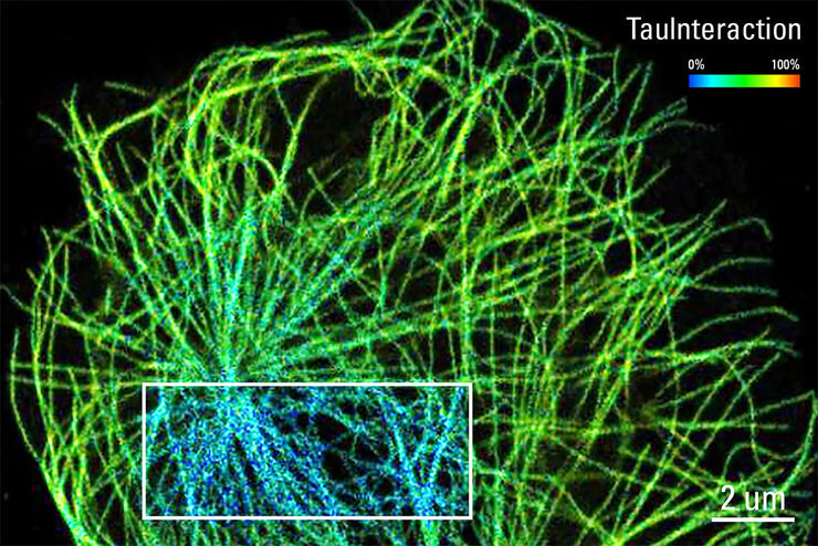
TauInteraction – Studying Molecular Interactions with TauSense
Fluorescence microscopy constitutes one of the pillars in life sciences and is a tool commonly used to unveil cellular structure and function. A key advantage of fluorescence microscopy resides in the…
Loading...

Studying Wound Healing of Smooth Muscle Cells
This article discusses how wound healing of cultured smooth muscle cells (SMCs) in multiwell plates can be reliably studied over time with less effort using a specially configured Leica inverted…
