
Science Lab
Science Lab
Das Wissensportal von Leica Microsystems bietet Ihnen Wissens- und Lehrmaterial zu den Themen der Mikroskopie. Die Inhalte sind so konzipiert, dass sie Einsteiger, erfahrene Praktiker und Wissenschaftler gleichermaßen bei ihrem alltäglichen Vorgehen und Experimenten unterstützen. Entdecken Sie interaktive Tutorials und Anwendungsberichte, erfahren Sie mehr über die Grundlagen der Mikroskopie und High-End-Technologien - werden Sie Teil der Science Lab Community und teilen Sie Ihr Wissen!
Filter articles
Tags
Berichtstyp
Produkte
Loading...

Mapping Tumor Immune Landscape with AI-Powered Spatial Proteomics
Spatial mapping of untreated tumors provides an overview of the tumor immune architecture, useful for understanding therapeutic responses. Immunocompetent murine models are essential for identifying…
Loading...

Virologie
Liegt Ihr Forschungsschwerpunkt auf Virusinfektionen und -krankheiten? Erfahren Sie, wie Sie mit Lösungen für Bildgebung und Probenvorbereitung von Leica Microsystems mehr Erkenntnisse in der…
Loading...

Kryo-Elektronen-Tomographie
Mit der Kryo-Elektronentomographie (CryoET) lassen sich Biomoleküle in ihrer zellulären Umgebung mit einer noch nie dagewesenen Auflösung von weniger als einem Nanometer auflösen.
Loading...

Zebrafisch-Forschung
Für optimale Ergebnisse während der Bewertung, Sortierung, Manipulation und Bildgebung von Modellorganismen ist es entscheidend feine Details und Strukturen genauestens zu erkennen. Das bildet die…
Loading...
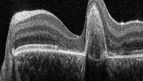
A Guide to OCT
Leica Optical Coherence Tomography (OCT) systems support ophthalmologists, ophthalmic surgeons, and researchers with easy-to-use, high-quality imaging technology.
Loading...

Aneurysm Clipping: Assessing Perforators in Real-time with AR Fluorescence
This article covers two aneurysm clipping cases highlighting the clinical benefits of GLOW800 Augmented Reality Fluorescence application in neurosurgery, based on insights from Prof. Tohru Mizutani,…
Loading...

How does Real-time OCT Imaging Impact Precision in Corneal Surgery?
Corneal surgery is a highly specialized field. It requires great surgical precision to overcome challenges such as visualizing clearly the full anterior chamber, performing Descemet membrane peeling…
Loading...

How to Achieve Brain Tissue Resection with GLOW400 AR
Intraoperative MRI is one form of real-time intraoperative visualization, but if more in-depth visualization to identify a tumor during surgery is wanted, intraoperative fluorescence diagnostics is…
Loading...

How to Automatically Obtain Fluorescent Cells of Interest in a Block-face
Block-face created by automatic trimming under fluorescence.
Mammalian cells of interest, stained with CellTrackerTM Green are visualized within the block-face using the UC Enuity equipped with the…
