
Science Lab
Science Lab
Das Wissensportal von Leica Microsystems bietet Ihnen Wissens- und Lehrmaterial zu den Themen der Mikroskopie. Die Inhalte sind so konzipiert, dass sie Einsteiger, erfahrene Praktiker und Wissenschaftler gleichermaßen bei ihrem alltäglichen Vorgehen und Experimenten unterstützen. Entdecken Sie interaktive Tutorials und Anwendungsberichte, erfahren Sie mehr über die Grundlagen der Mikroskopie und High-End-Technologien - werden Sie Teil der Science Lab Community und teilen Sie Ihr Wissen!
Filter articles
Tags
Berichtstyp
Produkte
Loading...

Modellorganismen in der Forschung
Modellorganismen sind Spezies, mit denen Forscher bestimmte biologische Vorgänge untersuchen. Sie haben genetische Ähnlichkeiten mit Menschen und werden häufig in Forschungsbereichen wie Genetik,…
Loading...

Virologie
Liegt Ihr Forschungsschwerpunkt auf Virusinfektionen und -krankheiten? Erfahren Sie, wie Sie mit Lösungen für Bildgebung und Probenvorbereitung von Leica Microsystems mehr Erkenntnisse in der…
Loading...

Wie die Analyse von Meeresmikroorganismen durch Hochdruckgefrieren verbessert werden kann
Die ultrastrukturelle Analyse von Umweltproben, hier Dinoflagellaten, bleibt heutzutage eine Herausforderung. Hier zeigen wir, dass die Durchführung von Hochdruckgefrieren (HPF) vor Ort die…
Loading...

How to Successfully Perform Live-cell CLEM
The Leica Nano workflow provides a streamlined live-cell CLEM solution for getting insight bout structural changes of cellular components over time. Besides the technical handling described in the…
Loading...

How to Successfully Implement Coral Life
The live-cell CLEM workflow allows you to capture dynamic information related to a relevant biological process as it happens and put these observations into their ultrastructural context. The Leica…
Loading...

How to Improve Live Cell Imaging with Coral Life
For live-cell CLEM applications, light microscopy imaging is a critical step for identifying the right cell in the right state at the right time. In this article, Leica experts share their insights on…
Loading...

How to Keep Your Samples Under Physiological Conditions
The Coral Life workflow combines dynamic data with the best possible sample fixation by high pressure freezing. However, good sample preservation won’t help if your cells are stressed by temperature…
Loading...
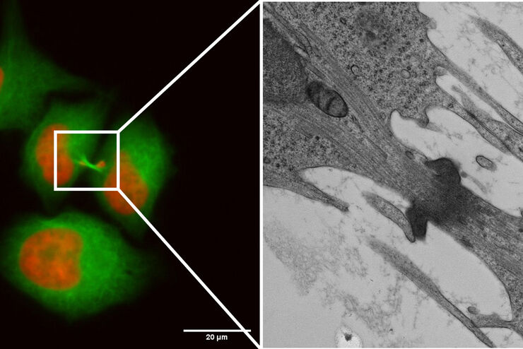
Capture life as it happens
With the Leica Nano Workflow, searching for the needle in the haystack is a thing of the past. Take advantage of correlative light and electron microscopy to identify directly the right cell at the…
Loading...
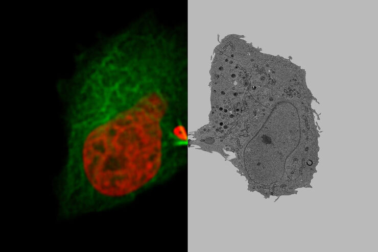
Putting Dynamic Live Cell Data into the Ultrastructural Context
With workflow Coral Life, searching for a needle in the haystack is a thing of the past. Take advantage of correlative light and electron microscopy to identify directly the right cell at the right…
