
Science Lab
Science Lab
Das Wissensportal von Leica Microsystems bietet Ihnen Wissens- und Lehrmaterial zu den Themen der Mikroskopie. Die Inhalte sind so konzipiert, dass sie Einsteiger, erfahrene Praktiker und Wissenschaftler gleichermaßen bei ihrem alltäglichen Vorgehen und Experimenten unterstützen. Entdecken Sie interaktive Tutorials und Anwendungsberichte, erfahren Sie mehr über die Grundlagen der Mikroskopie und High-End-Technologien - werden Sie Teil der Science Lab Community und teilen Sie Ihr Wissen!
Filter articles
Tags
Berichtstyp
Produkte
Loading...
, SPY-Actin (cyan), and SiR-Tubulin (magenta). Instant Computational Clearing (ICC) was applied.")
How to Perform Dynamic Multicolor Time-Lapse Imaging
Live-cell imaging sheds light on diverse cellular events. As many of these events have fast dynamics, the microscope imaging system must be fast enough to record every detail. One major advantage of…
Loading...
 and CellEvent™ (yellow).")
Following Multiple Events during Staurosporine Apoptosis
In this video on demand, we show how adding additional markers to an apoptosis kit can markedly increase the amount of information a researcher can obtain from the same experiment. The simultaneous…
Loading...
, the cis-golgi matrix protein GM130 (AF488, green), and the trans-golgi network membrane protein TGN46 (AF647, red).")
Golgi Organizational Changes in Response to Cell Stress
In this video on demand, our special guest George Galea from EMBL Heidelberg will look at HeLa Kyoto cells treated with various chemotherapeutic agents to investigate their effect on the Golgi complex…
Loading...
 stained to show the nucleus")
3D Spatial Analysis Using Mica's AI-Enabled Microscopy Software
This video offers practical advice on the extraction of publication grade insights from microscopy images. Our special guest Luciano Lucas (Leica Microsystems) will illustrate how Mica’s AI-enabled…
Loading...

3D Tissue Imaging: From Fast Overview To High Resolution With One Click
3D Tissue imaging is a widespread discipline in the life sciences. Researchers use it to reveal detailed information of tissue composition and integrity, to make conclusions from experimental…
Loading...

How To Perform Fast & Stable Multicolor Live-Cell Imaging
With the help of live-cell imaging researchers gain insights into dynamic processes of living cells up to whole organisms. This includes intracellular as well as intercellular activities. Protein or…
Loading...

Imaging of Cardiac Tissue Regeneration in Zebrafish
Learn how to image cardiac tissue regeneration in zebrafish focusing on cell proliferation and response during recovery with Laura Peces-Barba Castaño from the Max Planck Institute.
Loading...
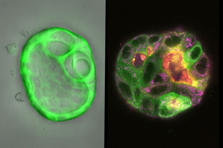
How Does The Cytoskeleton Transport Molecules?
VIDEO ON DEMAND - See how 3D cysts derived from MDCK cells help scientists understand how proteins are transported and recycled in tissues and the role of the cytoskeleton in this transport.
Loading...
 embryo, from sphere stage to somite stages.")
Studying Early Phase Development of Zebrafish Embryos
This video on demand focuses on combining widefield and confocal imaging to study the early-stage development of zebrafish embryos (Danio rerio), from oocyte to multicellular stage.
