
Science Lab
Science Lab
Das Wissensportal von Leica Microsystems bietet Ihnen Wissens- und Lehrmaterial zu den Themen der Mikroskopie. Die Inhalte sind so konzipiert, dass sie Einsteiger, erfahrene Praktiker und Wissenschaftler gleichermaßen bei ihrem alltäglichen Vorgehen und Experimenten unterstützen. Entdecken Sie interaktive Tutorials und Anwendungsberichte, erfahren Sie mehr über die Grundlagen der Mikroskopie und High-End-Technologien - werden Sie Teil der Science Lab Community und teilen Sie Ihr Wissen!
Filter articles
Tags
Berichtstyp
Produkte
Loading...

Applying AI and Machine Learning in Microscopy and Image Analysis
Prof. Emma Lundberg is a professor in cell biology proteomics at KTH Royal Institute of Technology, Sweden. She is also the director of the Cell Atlas, an integral part of the Swedish-based Human…
Loading...
, Astrocytes (GFAP, red), Nuclei (DAPI, blue).")
Multicolor Microscopy: The Importance of Multiplexing
The term multiplexing refers to the use of multiple fluorescent dyes to examine various elements within a sample. Multiplexing allows related components and processes to be observed in parallel,…
Loading...

A New Method for Convenient and Efficient Multicolor Imaging
The technique combining hyperspectral unmixing and phasor analysis was developed to simplify the process of getting images from a sample labeled with multiple fluorophores. This aggregate method…
Loading...

Considerations for Multiplex Live Cell Imaging
Simultaneous multicolor imaging for successful experiments: Live-cell imaging experiments are key to understand dynamic processes. They allow us to visually record cells in their living state, without…
Loading...
 and astrocytes (green) in a cortical spheroid derived from human induced pluripotent stem cells.")
Neuroscience Images
Neuroscience commonly uses microscopy to study the nervous system’s function and understand neurodegenerative diseases.
Loading...
 and astrocytes (green) in a cortical spheroid derived from human induced pluripotent stem cells.")
Download The Guide to Live Cell Imaging
In life science research, live cell imaging is an indispensable tool to visualize cells in a state as in vivo as possible. This E-book reviews a wide range of important considerations to take to…
Loading...

Introduction to Widefield Microscopy
This article gives an introduction to widefield microscopy, one of the most basic and commonly used microscopy techniques. It also shows the basic differences between widefield and confocal…
Loading...
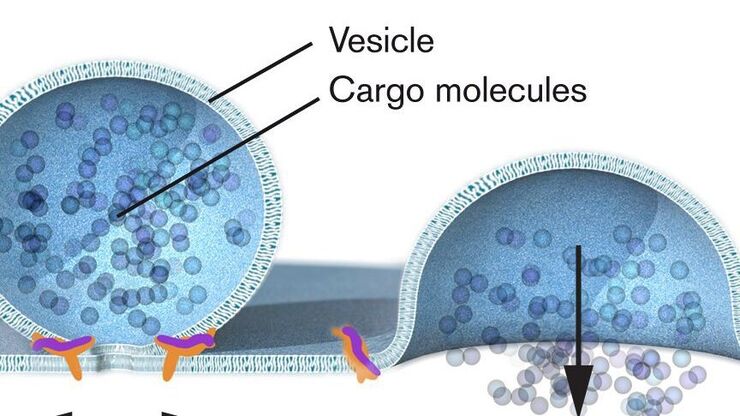
Nobel Prize 2013 in Physiology or Medicine for Discoveries of the Machinery Regulating Vesicle Traffic
On October 7th 2013, The Nobel Assembly at Karolinska Institutet has decided to award The Nobel Prize in Physiology or Medicine 2012 jointly to James E. Rothman, Randy W. Schekman and Thomas C. Südhof…
Loading...

Nobel Prize 2012 in Physiology or Medicine for Stem Cell Research
The Nobel Prize recognizes two scientists who discovered that mature, specialised cells can be reprogrammed to become immature cells capable of developing into all tissues of the body. Their findings…
