
Science Lab
Science Lab
Das Wissensportal von Leica Microsystems bietet Ihnen Wissens- und Lehrmaterial zu den Themen der Mikroskopie. Die Inhalte sind so konzipiert, dass sie Einsteiger, erfahrene Praktiker und Wissenschaftler gleichermaßen bei ihrem alltäglichen Vorgehen und Experimenten unterstützen. Entdecken Sie interaktive Tutorials und Anwendungsberichte, erfahren Sie mehr über die Grundlagen der Mikroskopie und High-End-Technologien - werden Sie Teil der Science Lab Community und teilen Sie Ihr Wissen!
Filter articles
Tags
Berichtstyp
Produkte
Loading...

Virologie
Liegt Ihr Forschungsschwerpunkt auf Virusinfektionen und -krankheiten? Erfahren Sie, wie Sie mit Lösungen für Bildgebung und Probenvorbereitung von Leica Microsystems mehr Erkenntnisse in der…
Loading...
. Courtesy: Thomas Mathivet, PhD")
Windows on Neurovascular Pathologies
Discover how innate immunity can sustain deleterious effects following neurovascular pathologies and the technological developments enabling longitudinal studies into these events.
Loading...
 and labelled with MitoTracker Green.")
The Power of Reproducibility, Collaboration and New Imaging Technologies
In this webinar you willl learn what impacts reproducibility in microscopy, what resources and initiatives there are to improve education and rigor and reproducibility in microscopy and how…
Loading...

Live-Cell Fluorescence Lifetime Multiplexing Using Organic Fluorophores
On-demand video: Imaging more subcellular targets by using fluorescence lifetime multiplexing combined with spectrally resolved detection.
Loading...
 und Akzeptormolekül (A), die am FRET (Förster-Resonanz-Energie-Transfer) teilnehmen.")
Was ist FRET mit FLIM (FLIM-FRET)?
Der Beitrag erläutert die FLIM-FRET-Methode, die Resonanzenergietransfer und Fluoreszenz-Lebensdauer-Imaging zur Untersuchung von Protein-Protein Wechselwirkungen kombiniert.
Loading...
Visualizing Protein-Protein Interactions by Non-Fitting and Easy FRET-FLIM Approaches
The Webinar with Dr. Sergi Padilla-Parra is about visualizing protein-protein interaction. He gives insight into non-fitting and easy FRET-FLIM approaches.
Loading...
 in 14 x 18 tiles. Lifetime gives an additional contrast that allows to differentiate different structures in histological stainings.")
Leitfaden zur Fluoreszenzlebensdauer-Imaging-Mikroskopie (FLIM)
Die Fluoreszenzlebensdauer ist ein Maß dafür, wie lange ein Fluorophor im Durchschnitt in seinem angeregten Zustand verbleibt, bevor er durch Aussendung eines Fluoreszenzphotons in den Grundzustand…
Loading...

Find Relevant Specimen Details from Overviews
Switch from searching image by image to seeing the full overview of samples quickly and identifying the important specimen details instantly with confocal microscopy. Use that knowledge to set up…
Loading...
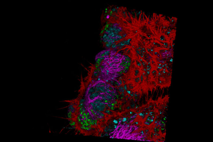
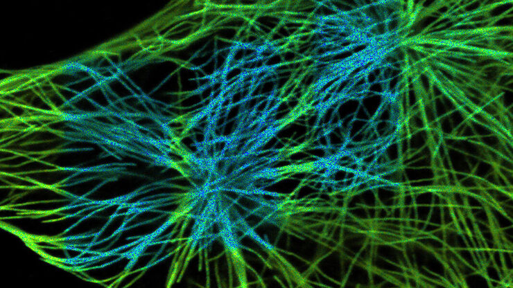
Fluorescence Lifetime-based Imaging Gallery
Confocal microscopy relies on the effective excitation of fluorescence probes and the efficient collection of photons emitted from the fluorescence process. One aspect of fluorescence is the emission…
