
Science Lab
Science Lab
Das Wissensportal von Leica Microsystems bietet Ihnen Wissens- und Lehrmaterial zu den Themen der Mikroskopie. Die Inhalte sind so konzipiert, dass sie Einsteiger, erfahrene Praktiker und Wissenschaftler gleichermaßen bei ihrem alltäglichen Vorgehen und Experimenten unterstützen. Entdecken Sie interaktive Tutorials und Anwendungsberichte, erfahren Sie mehr über die Grundlagen der Mikroskopie und High-End-Technologien - werden Sie Teil der Science Lab Community und teilen Sie Ihr Wissen!
Filter articles
Tags
Berichtstyp
Produkte
Loading...
 which was imaged with extended depth of field (EDOF) using digital microscopy.")
Depth of Field in Microscope Images
For microscopy imaging, depth of field is an important parameter when needing sharp images of sample areas with structures having significant changes in depth. In practice, depth of field is…
Loading...

What is Empty Magnification and How can Users Avoid it
The phenomenon of “empty magnification”, which can occur while using an optical, light, or digital microscope, and how it can be avoided is explained in this article. The performance of an optical…
Loading...
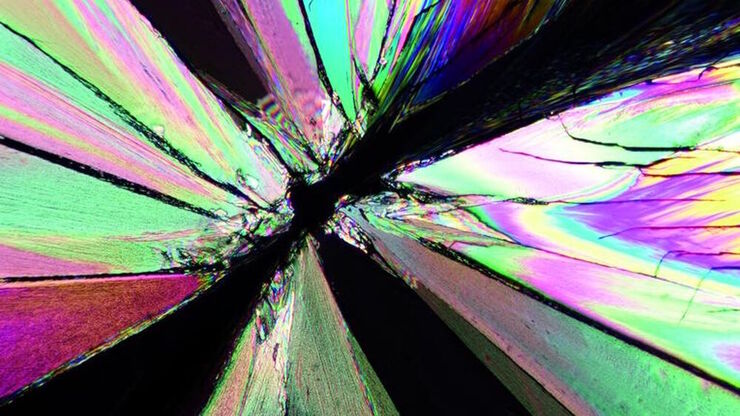
Das Prinzip der Polarisationsmikroskopie
Die Polarisationsmikroskopie wird in den Material- und Geowissenschaften routinemäßig eingesetzt, um Materialien und Mineralien anhand ihrer charakteristischen Brechungseigenschaften und Farben zu…
Loading...

Technical Terms for Digital Microscope Cameras and Image Analysis
Learn more about the basic principles behind digital microscope camera technologies, how digital cameras work, and take advantage of a reference list of technical terms from this article.
Loading...

Understanding Clearly the Magnification of Microscopy
To help users better understand the magnification of microscopy and how to determine the useful range of magnification values for digital microscopes, this article provides helpful guidelines.
Loading...

ISO 9022 Standard Part 11 - Testing Microscopes with Severe Conditions
This article describes a test to determine the robustness of Leica microscopes to mold and fungus growth. The test follows the specifications of the ISO 9022 part 11 standard for optical instruments.
Loading...

Life-Science-Forschung: Welche Mikroskopkamera ist die richtige für Sie?
Wie Sie entscheiden, welche Kamera für Ihre Life-Science-Mikroskopie-Experimente die Richtige ist. Welche Kamera von Leica Microsystems ist für Sie am besten geeignet?
Loading...

An Introduction to Fluorescence
This article gives an introduction to fluorescence and photoluminescence, which includes phosphorescence, explains the basic theory behind them, and how fluorescence is used for microscopy.
Loading...

Differential Interference Contrast (DIC) Microscopy
This article demonstrates how differential interference contrast (DIC) can be actually better than brightfield illumination when using microscopy to image unstained biological specimens.
