
Science Lab
Science Lab
Das Wissensportal von Leica Microsystems bietet Ihnen Wissens- und Lehrmaterial zu den Themen der Mikroskopie. Die Inhalte sind so konzipiert, dass sie Einsteiger, erfahrene Praktiker und Wissenschaftler gleichermaßen bei ihrem alltäglichen Vorgehen und Experimenten unterstützen. Entdecken Sie interaktive Tutorials und Anwendungsberichte, erfahren Sie mehr über die Grundlagen der Mikroskopie und High-End-Technologien - werden Sie Teil der Science Lab Community und teilen Sie Ihr Wissen!
Filter articles
Tags
Berichtstyp
Produkte
Loading...

Imaging of Anti-Cancer Drug Uptake in Spheroids using DLS
Spheroid 3D cell culture models mimic the physiology and functions of living tissues making them a useful tool to study tumor morphology and screen anti-cancer drugs. The drug AZD2014 is a recognized…
Loading...

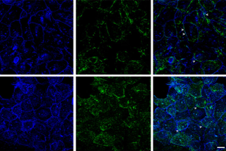
Designing your Research Study with Multiplexed IF Imaging
Multiplexed tissue analysis is a powerful technique that allows comparisons of cell-type locations and cell-type interactions within a single fixed tissue sample. It is common for researchers to ask…
Loading...

Be Confident in your Results with Cell DIVE Validated Antibodies
The Cell DIVE System includes a carefully curated list of hundreds of commercially available antibodies validated to offer optimal specificity and sensitivity in multiplexed imaging. That validation…
Loading...

Optimizing THUNDER Platform for High-Content Slide Scanning
With rising demand for full-tissue imaging and the need for FL signal quantitation in diverse biological specimens, the limits on HC imaging technology are tested, while user trainability and…
Loading...

Cancer Research Image Gallery
Fluorescence microscopy allows the study of changes occurring in tissue and cells during cancer development and progression. Techniques such as live cell imaging are critical to understand cancer…
Loading...

Regulators of Actin Cytoskeletal Regulation and Cell Migration in Human NK Cells
Dr. Mace will describe new advances in our understanding of the regulation of human NK cell actin cytoskeletal remodeling in cell migration and immune synapse formation derived from confocal and…
Loading...
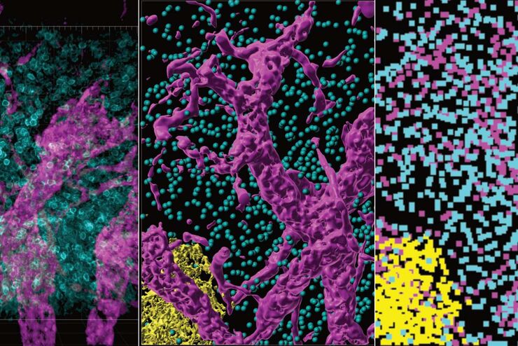
Improvement of Imaging Techniques to Understand Organelle Membrane Cell Dynamics
Understanding cell functions in normal and tumorous tissue is a key factor in advancing research of potential treatment strategies and understanding why some treatments might fail. Single-cell…
Loading...
From Organs to Tissues to Cells: Analyzing 3D Specimens with Widefield Microscopy
Obtaining high-quality data and images from thick 3D samples is challenging using traditional widefield microscopy because of the contribution of out-of-focus light. In this webinar, Falco Krüger…
Loading...
 stereo microscope for a task like surgery.")
Rodent and Small-Animal Surgery
Learn how you can perform rodent (mouse, rat, hamster) and small-animal surgery efficiently with a microscope for developmental biology and medical research applications by reading this article.
