
Science Lab
Science Lab
Das Wissensportal von Leica Microsystems bietet Ihnen Wissens- und Lehrmaterial zu den Themen der Mikroskopie. Die Inhalte sind so konzipiert, dass sie Einsteiger, erfahrene Praktiker und Wissenschaftler gleichermaßen bei ihrem alltäglichen Vorgehen und Experimenten unterstützen. Entdecken Sie interaktive Tutorials und Anwendungsberichte, erfahren Sie mehr über die Grundlagen der Mikroskopie und High-End-Technologien - werden Sie Teil der Science Lab Community und teilen Sie Ihr Wissen!
Filter articles
Tags
Berichtstyp
Produkte
Loading...
Enhancing Neurosurgery Teaching
Learn about the Serious Game in Intraoperative Neurosurgery and how it supports neurosurgical teaching and the acquisition of decision-making skills.
Loading...

How Intraoperative OCT Helps Gain Greater Insight in Glaucoma Surgery
Learn about the use of intraoperative Optical Coherence Tomography in glaucoma surgery and how it helps see subsurface tissue details.
Loading...

Ophthalmology: Visualization in Complex Cataract Surgery
Learn about the use of intraoperative Optical Coherence Tomography in cataract surgery and how it supports both standard and complex cataract surgery cases.
Loading...

Launching a Neurosurgical Department with Limited Resources
Learn about Dr. Claire Karekezi’s journey and experience launching a neurosurgical department within the Rwanda Military Hospital with limited resources.
Loading...

Use of AR Fluorescence in Neurovascular Surgery
Learn about the use of GLOW800 Augmented Reality in neurovascular surgery through clinical cases and videos, including aneurysm and tumor resection cases.
Loading...
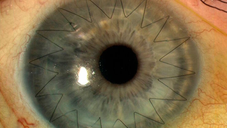
Ophthalmology Case Study: Corneal Transplantation
Learn about the use of intraoperative Optical Coherence Tomography in Corneal Transplantation and how it helps achieve correct positioning of donor tissue.
Loading...
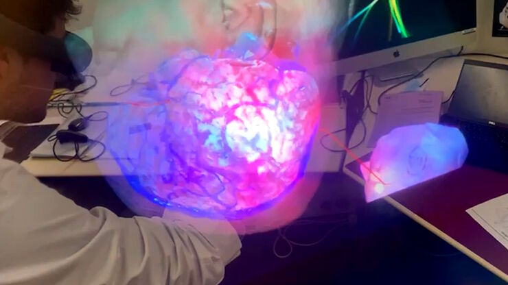
3D, AR & VR for Teaching in Neurosurgery
Discover the evolution of neurosurgical teaching and how 3D, Augmented Reality and Virtual Reality can help better learn anatomy and acquire surgical skills.
Loading...

Utility of Intraoperative OCT in Sub-Retinal Gene Therapy
Discover a case study on the use of intraoperative OCT for pediatric gene therapy and how it supports bleb placement and verifying the foveal contour.
Loading...
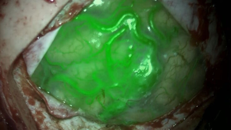
AR Fluorescence in Aneurysm Clipping and AVM Surgery
Discover how GLOW800 Augmented Reality fluorescence supports neurovascular surgical procedures and in particular aneurysm clipping and AVM surgery.
