
Science Lab
Science Lab
Das Wissensportal von Leica Microsystems bietet Ihnen Wissens- und Lehrmaterial zu den Themen der Mikroskopie. Die Inhalte sind so konzipiert, dass sie Einsteiger, erfahrene Praktiker und Wissenschaftler gleichermaßen bei ihrem alltäglichen Vorgehen und Experimenten unterstützen. Entdecken Sie interaktive Tutorials und Anwendungsberichte, erfahren Sie mehr über die Grundlagen der Mikroskopie und High-End-Technologien - werden Sie Teil der Science Lab Community und teilen Sie Ihr Wissen!
Filter articles
Tags
Berichtstyp
Produkte
Loading...

Targeting Active Recycling Nuclear Pore Complexes using Cryo Confocal Microscopy
In this article, how cryo light microscopy and, in particular cryo confocal microscopy, is used to improve the reliability of cryo EM workflows is described. The quality of the EM grids and samples is…
Loading...

How FLIM Microscopy Helps to Detect Microplastic Pollution
The use of autofluorescence in biological samples is a widely used method to gain detailed knowledge about systems or organisms. This property is not only found in biological systems, but also…
Loading...
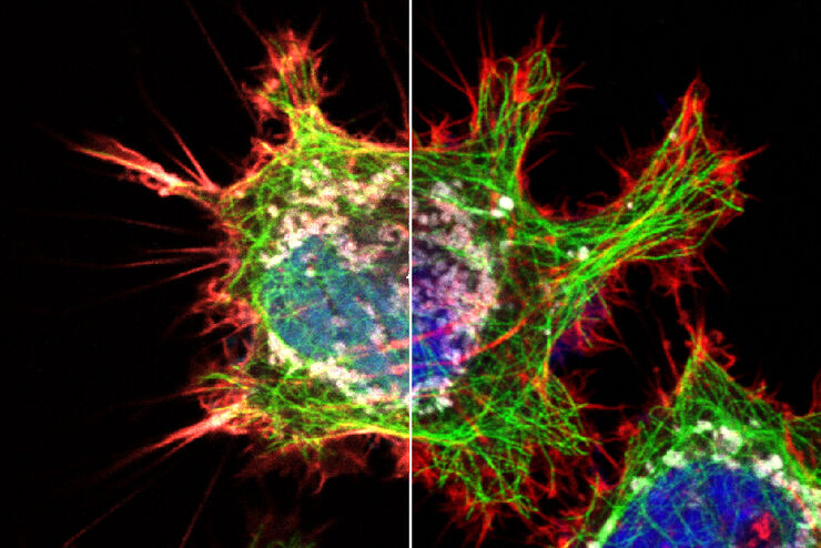
 - THUNDER Imager 3D Cell Culture Influenca virus – red, cilia – green, Nuclei – blue.")
How Can Immunofluorescence Aid Virology Research?
Modern virology research has become as crucial now as ever before due to the global COVID-19 pandemic. There are many powerful technologies and assays that virologists can apply to their research into…
Loading...
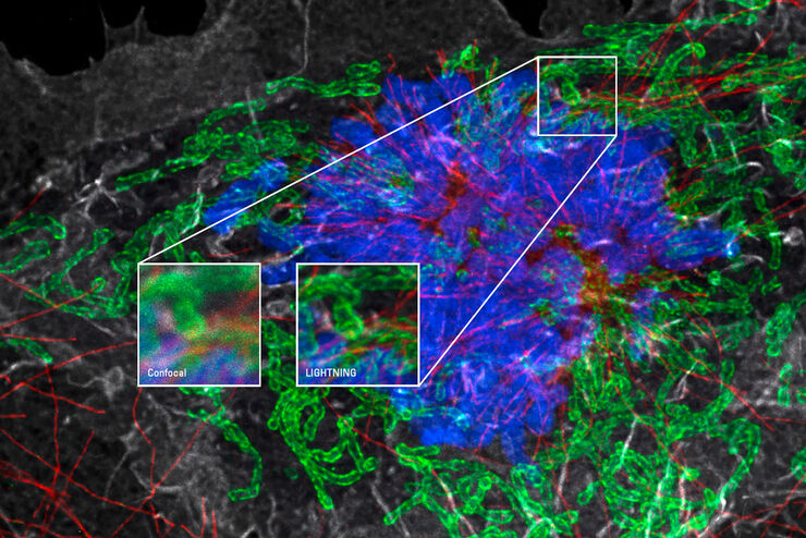
Mit LIGHTNING das Maximum an Informationen aus Ihrer Probe erhalten
LIGHTNING ist ein adaptiver Prozess zur Extraktion von Bildinformationen, bei dem vollautomatisch anderweitig nicht sichtbare Strukturen und feine Details sichtbar gemacht werden. Im Gegensatz zu…
Loading...

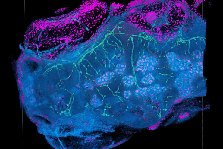
Microscopy in Virology
The coronavirus SARS-CoV-2, causing the Covid-19 disease effects our world in all aspects. Research to find immunization and treatment methods, in other words to fight this virus, gained highest…
Loading...
Explore Innovative Techniques to Separate Fluorophores with Overlapping Spectra
In this article we explore several strategies you can take to improve the separation of fluorophores and increase the number of fluorescent probes you can distinguish in your sample.
Loading...

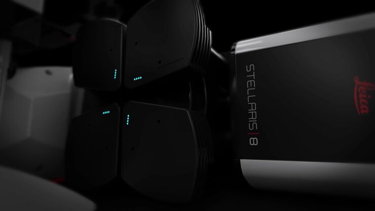
STELLARIS White Light Lasers
When it comes to choosing fluorescent probes for your multi-color experiments, you shouldn’t have to compromise. Now you can advance beyond conventional excitation sources that limit your fluorophore…
Loading...
TauSense Technology Imaging Tools
Leica Microsystems’ TauSense technology is a set of imaging modes based on fluorescence lifetime. Found at the core of the STELLARIS confocal platform, it will revolutionize your imaging experiments.…
Loading...
The Power HyD Detector Family
Powerful photon counting detectors on the STELLARIS confocal platform provide improved photon counting, ultra-sensitive imaging and more color options in the NIR spectrum.
