
Science Lab
Science Lab
Das Wissensportal von Leica Microsystems bietet Ihnen Wissens- und Lehrmaterial zu den Themen der Mikroskopie. Die Inhalte sind so konzipiert, dass sie Einsteiger, erfahrene Praktiker und Wissenschaftler gleichermaßen bei ihrem alltäglichen Vorgehen und Experimenten unterstützen. Entdecken Sie interaktive Tutorials und Anwendungsberichte, erfahren Sie mehr über die Grundlagen der Mikroskopie und High-End-Technologien - werden Sie Teil der Science Lab Community und teilen Sie Ihr Wissen!
Filter articles
Tags
Berichtstyp
Produkte
Loading...

A Guide to Darkfield Microscopes
A darkfield microscope offers a way to view the structures of many types of biological specimens in greater contrast without the need of stains.
Loading...
 and oblique (right) brightfield illumination using a Leica compound microscope. The defect on the wafer surface is clearly more visible with oblique illumination.")
Rapid Semiconductor Inspection with Microscope Contrast Methods
Semiconductor inspection during the production of patterned wafers and ICs (integrated circuits) is important for identifying and minimizing defects. To increase the efficiency of quality control in…
Loading...

Metallography – an Introduction
This article gives an overview of metallography and metallic alloy characterization. Different microscopy techniques are used to study the alloy microstructure, i.e., microscale structure of grains,…
Loading...

How to Adapt Grain Size Analysis of Metallic Alloys to Your Needs
Metallic alloys, such as steel and aluminum, have an important role in a variety of industries, including automotive and transportation. In this report, the importance of grain size analysis for alloy…
Loading...

Imaging and Analyzing Zebrafish, Medaka, and Xenopus
Discover how to image and analyze zebrafish, medaka, and Xenopus frog model organisms efficiently with a microscope for developmental biology applications from this article.
Loading...

Studying Caenorhabditis elegans (C. elegans)
Find out how you can image and study C. elegans roundworm model organisms efficiently with a microscope for developmental biology applications from this article.
Loading...
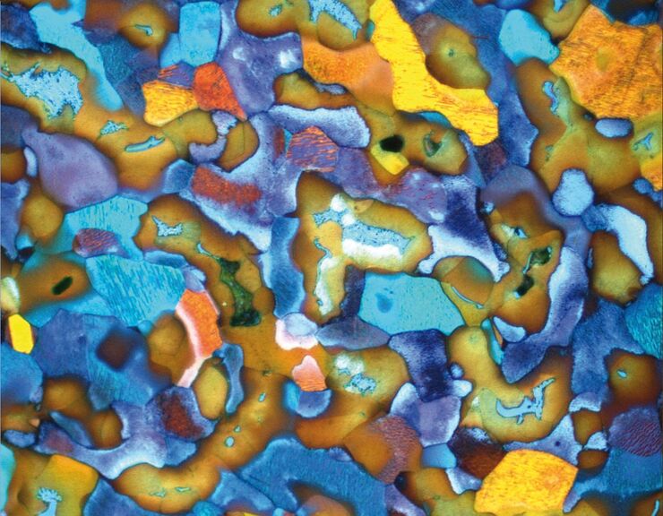
Metallography with Color and Contrast
The examination of microstructure morphology plays a decisive role in materials science and failure analysis. There are many possibilities of visualizing the real structures of materials in the light…
Loading...

Optical Contrast Methods
Optical contrast methods give the potential to easily examine living and colorless specimens. Different microscopic techniques aim to change phase shifts caused by the interaction of light with the…
