
Science Lab
Science Lab
Das Wissensportal von Leica Microsystems bietet Ihnen Wissens- und Lehrmaterial zu den Themen der Mikroskopie. Die Inhalte sind so konzipiert, dass sie Einsteiger, erfahrene Praktiker und Wissenschaftler gleichermaßen bei ihrem alltäglichen Vorgehen und Experimenten unterstützen. Entdecken Sie interaktive Tutorials und Anwendungsberichte, erfahren Sie mehr über die Grundlagen der Mikroskopie und High-End-Technologien - werden Sie Teil der Science Lab Community und teilen Sie Ihr Wissen!
Filter articles
Tags
Berichtstyp
Produkte
Loading...
A Guide to Super-Resolution
Find out more about Leica super-resolution microscopy solutions and how they can empower you to visualize in fine detail subcellular structures and dynamics.
Loading...

How to Study Gene Regulatory Networks in Embryonic Development
Join Dr. Andrea Boni by attending this on-demand webinar to explore how light-sheet microscopy revolutionizes developmental biology. This advanced imaging technique allows for high-speed, volumetric…
Loading...
.")
Dual-View LightSheet Microscope for Large Multicellular Systems
Visualizing the dynamics of complex multicellular systems is a fundamental goal in biology. To address the challenges of live imaging over large spatiotemporal scales, Franziska Moos et. al. present…
Loading...

Notable AI-based Solutions for Phenotypic Drug Screening
Learn about notable optical microscope solutions for phenotypic drug screening using 3D-cell culture, both planning and execution, from this free, on-demand webinar.
Loading...

Confocal Imaging of Immune Cells in Tissue Samples
In this webinar, you will discover how to perform 10-color acquisition using a confocal microscope. The challenges of imaged-based approaches to identify skin immune cells. A new pipeline to assess…
Loading...
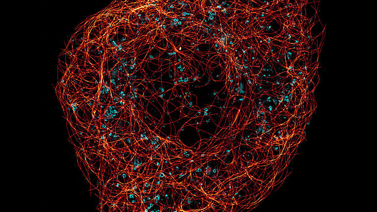
 and mito OM (red) in a live U2OS cell")
Multicolor 4D Super Resolution Light Sheet Microscopy
The AI Microscopy Symposium offers a unique forum for discussing the latest AI-based technologies and tools in the field of microscopy and biomedical imaging. In this scientific presentation, Yuxuan…
Loading...

Imaging of Anti-Cancer Drug Uptake in Spheroids using DLS
Spheroid 3D cell culture models mimic the physiology and functions of living tissues making them a useful tool to study tumor morphology and screen anti-cancer drugs. The drug AZD2014 is a recognized…
Loading...
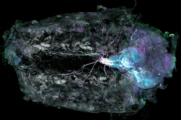
Understanding Motor Sequence Generation Across Spatiotemporal Scales
We have developed a microscopy-based pipeline to characterize a developmentally critical behavior at the pupal stage of development, called the ecdysis sequence. We study brain-wide neuronal activity…
Loading...
![3D glomeruli in a portion of an ECi-cleared kidney scanned by light sheet microscopy. Courtesy of Prof. Norbert Gretz, Medical Faculty Mannheim, University of Heidelberg [1].](/fileadmin/_processed_/d/d/csm_DLS-Sample-Preparation-Intr_915e0fd7c2.jpg "3D glomeruli in a portion of an ECi-cleared kidney scanned by light sheet microscopy. Courtesy of Prof. Norbert Gretz, Medical Faculty Mannheim, University of Heidelberg [1].")
Using Mounting Frames for Light Sheet Sample Preparation
Sample handling is an important topic in the context of Light Sheet Microscopy. The TCS SP8 DLS integrates Light Sheet technology into an inverted confocal platform and can hence make use of general…
