
Science Lab
Science Lab
Das Wissensportal von Leica Microsystems bietet Ihnen Wissens- und Lehrmaterial zu den Themen der Mikroskopie. Die Inhalte sind so konzipiert, dass sie Einsteiger, erfahrene Praktiker und Wissenschaftler gleichermaßen bei ihrem alltäglichen Vorgehen und Experimenten unterstützen. Entdecken Sie interaktive Tutorials und Anwendungsberichte, erfahren Sie mehr über die Grundlagen der Mikroskopie und High-End-Technologien - werden Sie Teil der Science Lab Community und teilen Sie Ihr Wissen!
Filter articles
Tags
Berichtstyp
Produkte
Loading...
Phasenkontrast
Mit einem Phasenkontrast-Lichtmikroskop können die Strukturen vieler Arten von biologischen Präparaten mit einem größeren Kontrast betrachtet werden, ohne dass die Probe eingefärbt werden muss.
Loading...
 mit Phasenkontrast.")
Phasenkontrast und Mikroskopie
Dieser Artikel erklärt den Phasenkontrast, eine optische Mikroskopietechnik, die feine Details von ungefärbten, transparenten Proben sichtbar macht, die mit gewöhnlicher Hellfeldbeleuchtung schwer zu…
Loading...

How to Determine Cell Confluency with a Digital Microscope
This article shows how to measure cell confluency in an easy and consistent way with Mateo TL, increasing confidence in downstream experiments.
Loading...

How to do a Proper Cell Culture Quick Check
In order to successfully work with mammalian cell lines, they must be grown under controlled conditions and require their own specific growth medium. In addition, to guarantee consistency their growth…
Loading...
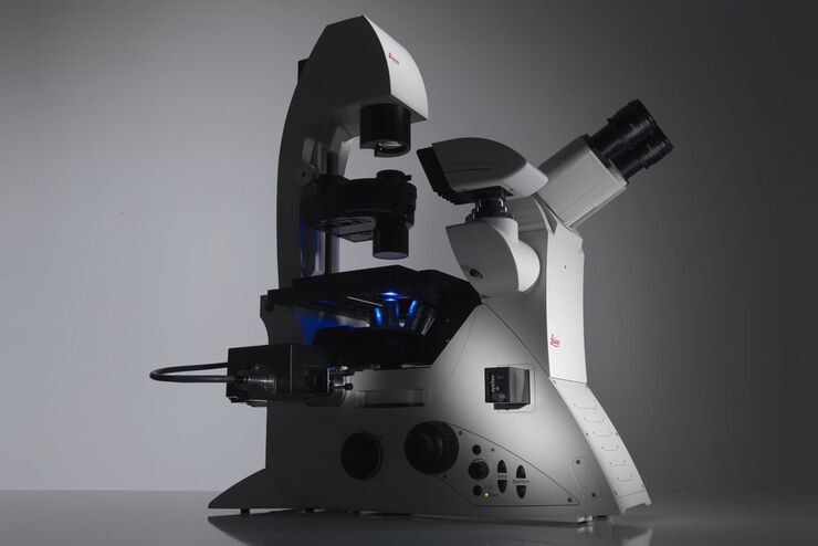
Factors to Consider When Selecting a Research Microscope
An optical microscope is often one of the central devices in a life-science research lab. It can be used for various applications which shed light on many scientific questions. Thereby the…
Loading...

Introduction to Mammalian Cell Culture
Mammalian cell culture is one of the basic pillars of life sciences. Without the ability to grow cells in the lab, the fast progress in disciplines like cell biology, immunology, or cancer research…
Loading...

Introduction to Widefield Microscopy
This article gives an introduction to widefield microscopy, one of the most basic and commonly used microscopy techniques. It also shows the basic differences between widefield and confocal…
Loading...
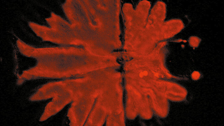
 inoculated with cowpea mosaic virus (CPMV) containing the GFP-gene inserted between the movement protein (MP) and the capsid proteins (CPs) in the viral RNA 2")
Introduction to Live-Cell Imaging
The understanding of complex and fast cellular dynamics is an important step to get insight into biological processes. Therefore, today’s life science research more and more demands studying…
Loading...

Optical Contrast Methods
Optical contrast methods give the potential to easily examine living and colorless specimens. Different microscopic techniques aim to change phase shifts caused by the interaction of light with the…
