
Science Lab
Science Lab
Das Wissensportal von Leica Microsystems bietet Ihnen Wissens- und Lehrmaterial zu den Themen der Mikroskopie. Die Inhalte sind so konzipiert, dass sie Einsteiger, erfahrene Praktiker und Wissenschaftler gleichermaßen bei ihrem alltäglichen Vorgehen und Experimenten unterstützen. Entdecken Sie interaktive Tutorials und Anwendungsberichte, erfahren Sie mehr über die Grundlagen der Mikroskopie und High-End-Technologien - werden Sie Teil der Science Lab Community und teilen Sie Ihr Wissen!
Filter articles
Tags
Berichtstyp
Produkte
Loading...

Improve 3D Cell Biology Workflow with Light Sheet Microscopy
Understanding the sub-cellular mechanisms in carcinogenesis is of crucial importance for cancer treatment. Popular cellular models comprise cancer cells grown as monolayers. But this approach…
Loading...
 and YOYO 1 iodide (Nucleus).")
Real Time Images of 3D Specimens with Sharp Contrast Free of Haze
THUNDER Imagers deliver in real time images of 3D specimens with sharp contrast, free of the haze or out-of-focus blur typical of widefield systems. They can even image clearly places deep inside a…
Loading...
Workflows & Protocols: How to Use a Leica Laser Microdissection System and Qiagen Kits for Successful RNA Analysis
Laser Microdissection (LMD) allows isolating individual cells or chromosomes and is a well established technique for sample preparation prior downstream analysis of the nucleic acid content via PCR or…
Loading...
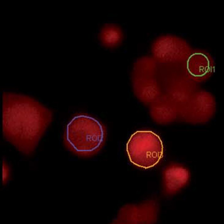
Widefield Calcium Imaging with Calcium Indicator Fura2
In eukaryotic cells Ca2+ is one of the most widespread second messengers used in signal transduction pathways. Intracellular levels of Ca2+ are usually kept low, as Ca2+ often forms insoluble…
