
Science Lab
Science Lab
Das Wissensportal von Leica Microsystems bietet Ihnen Wissens- und Lehrmaterial zu den Themen der Mikroskopie. Die Inhalte sind so konzipiert, dass sie Einsteiger, erfahrene Praktiker und Wissenschaftler gleichermaßen bei ihrem alltäglichen Vorgehen und Experimenten unterstützen. Entdecken Sie interaktive Tutorials und Anwendungsberichte, erfahren Sie mehr über die Grundlagen der Mikroskopie und High-End-Technologien - werden Sie Teil der Science Lab Community und teilen Sie Ihr Wissen!
Filter articles
Tags
Berichtstyp
Produkte
Loading...

Collecting Light: The Importance of Numerical Aperture in Microscopy
Numerical aperture (abbreviated as ‘NA’) is an important consideration when trying to distinguish detail in a specimen viewed down the microscope. NA is a number without units and is related to the…
Loading...

Laser Beam Shaping for Multicolor Multiphoton Microscopy
Multiphoton Microscopy is one of the current hot topics in life science research. The new Leica TCS SP8 DIVE from Leica Microsystems presents a series of beneficial new innovations, including a freely…
Loading...

Introduction to Widefield Microscopy
This article gives an introduction to widefield microscopy, one of the most basic and commonly used microscopy techniques. It also shows the basic differences between widefield and confocal…
Loading...
Optimization of the Interplay of Optical Components for Aberration Free Microscopy
Optical microscopes are used to magnify objects which are otherwise invisible for the human eye. For this purpose high quality optics is necessary to achieve appropriate resolution. However, besides…
Loading...
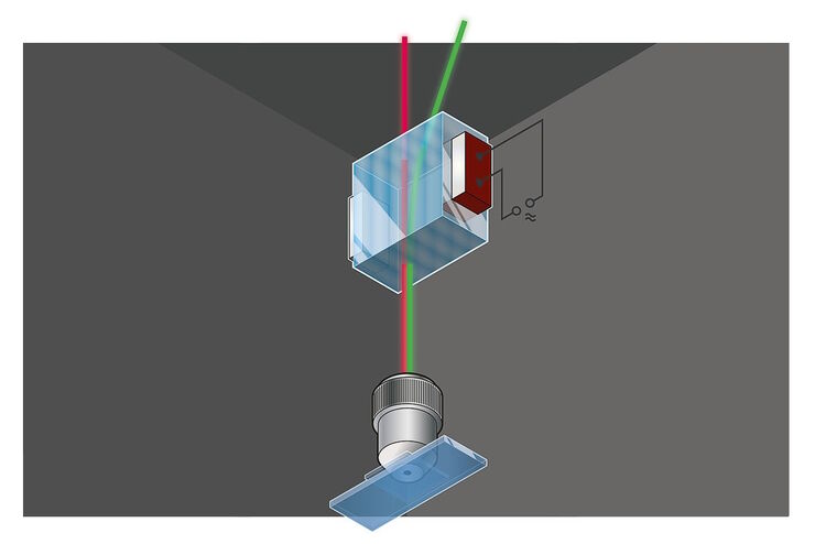
Primary Beam Splitting Devices for Confocal Microscopes
Current fluorescence microscopy employs incident illumination which requires separation of illumination and emission light. The classical device performing this separation is a color-dependent beam…
Loading...

Photoactivatable, Photoconvertible, and Photoswitchable Fluorescent Proteins
Fluorescent proteins (FPs) such as GFP, YFP or DsRed are powerful tools to visualize cellular components in living cells. Nevertheless, there are circumstances when classical FPs reach their limits.…
Loading...

Was macht das Pinhole im konfokalen Mikroskop?
Diese kurze einführende Schrift soll die Bedeutung des Pinholes erläutern und ist für Leser gedacht, die nicht allzu viel Zeit mit der Theorie und den Details der konfokalen Mikroskopie verbringen…
Loading...
 stereo microscope for a task like surgery.")
Rodent and Small-Animal Surgery
Learn how you can perform rodent (mouse, rat, hamster) and small-animal surgery efficiently with a microscope for developmental biology and medical research applications by reading this article.
Loading...

Navigating Through the Brain
One of the challenges of neurosurgery is orientation at the surgical site. When resecting tumors, removing arteriovenous malformations, or clipping aneurysms, surgeons often have to work near healthy…
