
Science Lab
Science Lab
Das Wissensportal von Leica Microsystems bietet Ihnen Wissens- und Lehrmaterial zu den Themen der Mikroskopie. Die Inhalte sind so konzipiert, dass sie Einsteiger, erfahrene Praktiker und Wissenschaftler gleichermaßen bei ihrem alltäglichen Vorgehen und Experimenten unterstützen. Entdecken Sie interaktive Tutorials und Anwendungsberichte, erfahren Sie mehr über die Grundlagen der Mikroskopie und High-End-Technologien - werden Sie Teil der Science Lab Community und teilen Sie Ihr Wissen!
Filter articles
Tags
Berichtstyp
Produkte
Loading...
BABB Clearing and Imaging for High Resolution Confocal Microscopy
Multipohoton microscopy experiment using Leica TCS SP8 MP and Leica 20x/0.95 NA BABB immersion objective.
Understanding kidney microanatomy is key to detecting and identifying early events in kidney…
Loading...
")
Epoxy Resin Embedding of Animal and Human Tissues for Pathological Diagnosis and Research
Application Note for Leica EM AMW - The tissues were fixed in the modified Karnovsky fixative generally by immersion overnight (at minimum 4h at room temperature). Then pieces of approx. 1mm3 were cut…
Loading...
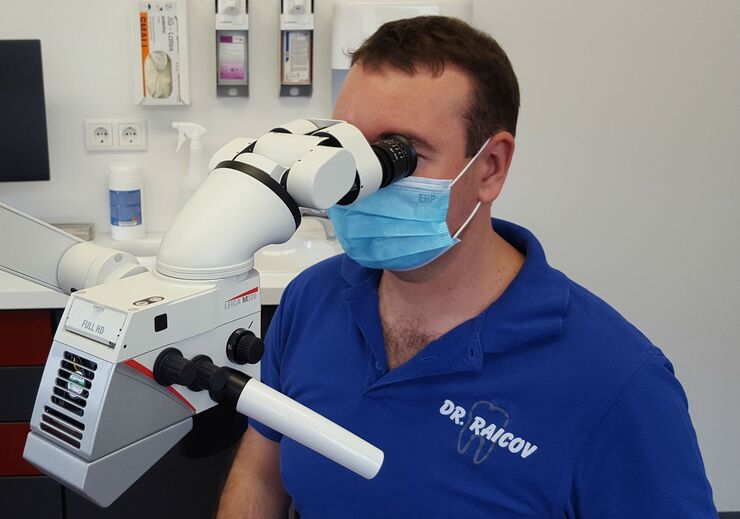
Das Dentalmikroskop in der Endodontie
In der Zahnmedizin unterstützt ein heller, vergrößerter Blick in tiefe Kavitäten die detaillierte Diagnose und präzise Therapie, insbesondere im Bereich der Endodontie. In diesem Interview erklärt Dr.…
Loading...

Human Blood Cells Protocol
Application Note for Leica EM CPD300 - Life Science Research. Species: Human (Homo sapiens)
Critical point drying of human blood with subsequent platinum / palladium coating and SEM analysis.
Loading...

Sechs wichtige Faktoren für die Auswahl eines Dentalmikroskops
In der Zahnmedizin ist das Operationsmikroskop mittlerweile ein wichtiges Werkzeug für qualitativ hochwertige und erfolgreiche Operationen, insbesondere im Bereich der Endodontie. Ein Mikroskop…
Loading...

Improvement of Metallic Thin Films for HR-SEM by Using DC Magnetron Sputter Coater
Preparation techniques, like several kinds of coating methods play an important role for high resolution scanning electron microscopy (HR-SEM). Nonconductive sample like biological and synthetic…
Loading...

Infinity Optical Systems
“Infinity Optics” refers to the concept of a beam path with parallel rays between the objective and the tube lens of a microscope. Flat optical components can be brought into this “Infinity Space”…
Loading...

Imaging of Host Cell-bacteria Interactions using Correlative Microscopy under Cryo-conditions
Pathogenic bacteria have developed intriguing strategies to establish and promote infections in their respective hosts. Most bacterial pathogens initiate infectious diseases by adhering to host cells…
Loading...

Cataract Surgery with CoAx4 Illumination
A stable red reflex is one of the most important features of an ophthalmic surgical microscope for cataract surgery. It’s the red reflex that makes the structure of the lens visible and thus makes for…
