
Science Lab
Science Lab
Das Wissensportal von Leica Microsystems bietet Ihnen Wissens- und Lehrmaterial zu den Themen der Mikroskopie. Die Inhalte sind so konzipiert, dass sie Einsteiger, erfahrene Praktiker und Wissenschaftler gleichermaßen bei ihrem alltäglichen Vorgehen und Experimenten unterstützen. Entdecken Sie interaktive Tutorials und Anwendungsberichte, erfahren Sie mehr über die Grundlagen der Mikroskopie und High-End-Technologien - werden Sie Teil der Science Lab Community und teilen Sie Ihr Wissen!
Filter articles
Tags
Berichtstyp
Produkte
Loading...

Workflows & Protocols: How to Isolate Individual Chromosomes with Laser Microdissection
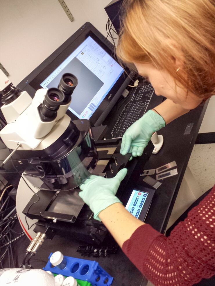
During the first Leica Workshop in Brazil, at the Centro de Energia Nuclear na Agricultura/USP (CENA), the participants learned how to prepare samples for laser microdissection (LMD) using a cryotome.…
Loading...
")
A Brief History of Light Microscopy
The history of microscopy begins in the Middle Ages. As far back as the 11th century, plano-convex lenses made of polished beryl were used in the Arab world as reading stones to magnify manuscripts.…
Loading...

Vom Licht zur Erleuchtung: Sensoren und Messverfahren in der konfokalen Mikroskopie
In diesem Beitrag werden die wichtigsten Sensoren kurz vorgestellt, die in der konfokalen Mikroskopie verwendet werden. Mit konfokaler Mikroskopie ist hier „True Confo-cal Scanning“ gemeint, also das…
Loading...
Workflows & Protocols: How to Use a Leica Laser Microdissection System and Qiagen Kits for Successful RNA Analysis
Laser Microdissection (LMD) allows isolating individual cells or chromosomes and is a well established technique for sample preparation prior downstream analysis of the nucleic acid content via PCR or…
Loading...

Confocal and Light Sheet Imaging
Optical imaging instrumentation can magnify tiny objects, zoom in on distant stars and reveal details that are invisible to the naked eye. But it notoriously suffers from an annoying problem: the…
Loading...

Universal PAINT – Dynamic Super-Resolution Microscopy
Super-resolution microscopy techniques have revolutionized biology for the last ten years. With their help cellular components can now be visualized at the size of a protein. Nevertheless, imaging…
Loading...
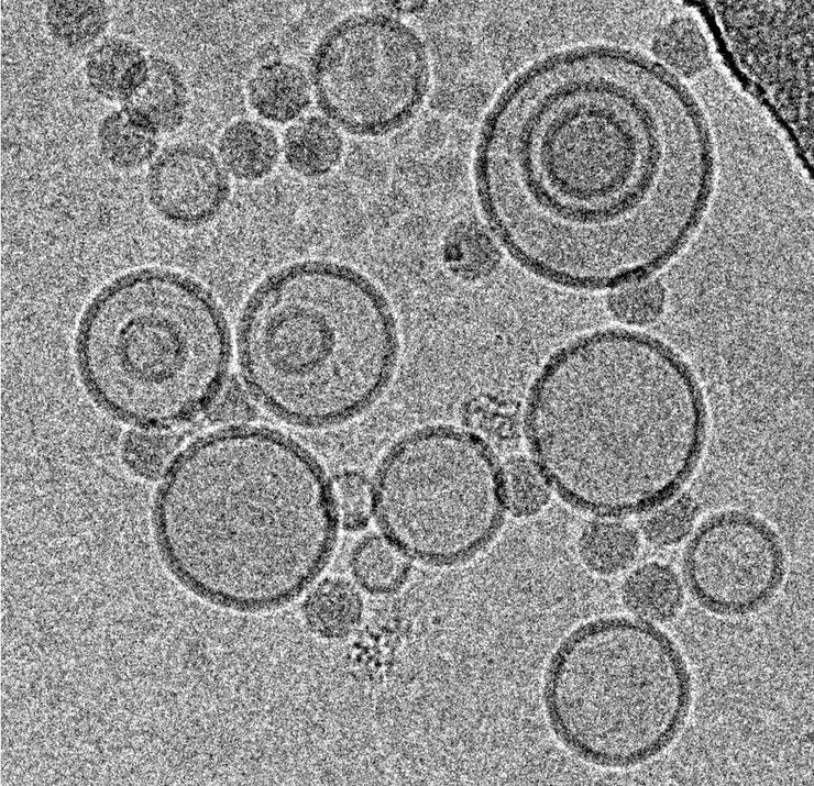
Immersion Freezing for Cryo-Transmission Electron Microscopy: Applications
A well established usage case for cryo-TEM is three-dimensional reconstruction of isolated macromolecules, virus particles, or filaments. On one hand, these approaches are based on averaging of…
Loading...
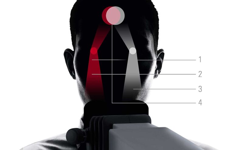
FusionOptics in Neurosurgery and Ophthalmology – for a Larger 3D Area in Focus
Neurosurgeons and ophthalmologists deal with delicate structures, deep or narow cavities and tiny structures with vitally important functions. A clear, three-dimensional view on the surgical field is…
Loading...
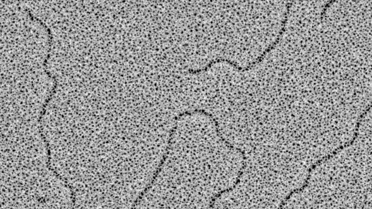
Glycerol Spraying/Platinum Low Angle Rotary Shadowing of DNA with the Leica EM ACE600 e-beam
Glycerol spraying/low angle rotary shadowing (Aebi and Baschong, 2006) is a preparation technique used in biology to visualize structures yielding insufficent contrast with other techniques, due to…
