
Science Lab
Science Lab
Das Wissensportal von Leica Microsystems bietet Ihnen Wissens- und Lehrmaterial zu den Themen der Mikroskopie. Die Inhalte sind so konzipiert, dass sie Einsteiger, erfahrene Praktiker und Wissenschaftler gleichermaßen bei ihrem alltäglichen Vorgehen und Experimenten unterstützen. Entdecken Sie interaktive Tutorials und Anwendungsberichte, erfahren Sie mehr über die Grundlagen der Mikroskopie und High-End-Technologien - werden Sie Teil der Science Lab Community und teilen Sie Ihr Wissen!
Filter articles
Tags
Berichtstyp
Produkte
Loading...
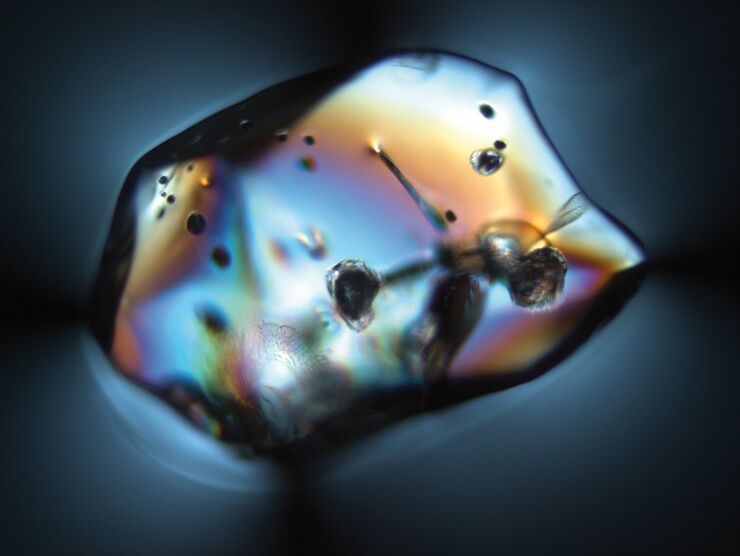
Quality as Clear as Glass - Polarizing Microscopy in Glass Production
An exquisite beverage deserves a high-quality glass. Even the ancient Romans made artistically crafted drinking glasses. In the Middle Ages, Venetian glassmakers were famous for the purity of their…
Loading...

The History of Stereo Microscopy
This article gives an overview on the history of stereo microscopes. The development and evolution from handcrafted instruments (late 16th to mid-18th century) to mass produced ones the last 150…
