
Science Lab
Science Lab
Das Wissensportal von Leica Microsystems bietet Ihnen Wissens- und Lehrmaterial zu den Themen der Mikroskopie. Die Inhalte sind so konzipiert, dass sie Einsteiger, erfahrene Praktiker und Wissenschaftler gleichermaßen bei ihrem alltäglichen Vorgehen und Experimenten unterstützen. Entdecken Sie interaktive Tutorials und Anwendungsberichte, erfahren Sie mehr über die Grundlagen der Mikroskopie und High-End-Technologien - werden Sie Teil der Science Lab Community und teilen Sie Ihr Wissen!
Filter articles
Tags
Berichtstyp
Produkte
Loading...

Quality Control Under the Microscope
Fast-rising demand for electric vehicles is one of the market’s main drivers, but there are other hotspots of growth, including the rise in renewable energy installations, such as photovoltaic panels,…
Loading...

Multiplexed Imaging Types, Benefits and Applications
Multiplexed imaging is an emerging and exciting way to extract information from human tissue samples by visualizing many more biomarkers than traditional microscopy. By observing many biomarkers…
Loading...

3D Tissue Imaging: From Fast Overview To High Resolution With One Click
3D Tissue imaging is a widespread discipline in the life sciences. Researchers use it to reveal detailed information of tissue composition and integrity, to make conclusions from experimental…
Loading...

How To Perform Fast & Stable Multicolor Live-Cell Imaging
With the help of live-cell imaging researchers gain insights into dynamic processes of living cells up to whole organisms. This includes intracellular as well as intercellular activities. Protein or…
Loading...
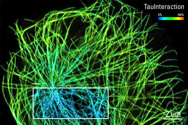
TauInteraction – Studying Molecular Interactions with TauSense
Fluorescence microscopy constitutes one of the pillars in life sciences and is a tool commonly used to unveil cellular structure and function. A key advantage of fluorescence microscopy resides in the…
Loading...

Technische Sauberkeit von Automobilkomponenten und -teilen
In diesem Artikel werden die ISO-Norm 16232 und die VDA 19-Richtlinien erläutert und die Verfahren zur Partikelanalyse kurz zusammengefasst. Diese liefern wichtige Kriterien für die Sauberkeit von…
Loading...

Verbesserte Ergonomie dank Dentalmikroskop
Dr. David Blanc, ein Zahnchirurg und Ergonomieberater, an einem Dentalmikroskop mit besonders niedrigen Binokulartuben. Dank optimierter Ergonomie hat Dr. Blanc einen höheren Bedienkomfort, da er bei…
Loading...
, unsaturated lipids (magenta, 3050 cm-1), collagen (SHG, cyan). Sample courtesy of R. Rudolf, J Klicks, Hochschule Mannheim")
The Potential of Coherent Raman Scattering Microscopy at a Glance
Coherent Raman scattering microscopy (CRS) is a powerful approach for label-free, chemically specific imaging. It is based on the characteristic intrinsic vibrational contrast of molecules in the…
Loading...

Simplifying Complex Fluorescence Multiwell Plate Assays
Apoptosis, or programmed cell death, occurs during organism embryo development to eliminate unwanted cells and during healing in adults to rid the body of damaged cells and help prevent cancer.…
