
Science Lab
Science Lab
Das Wissensportal von Leica Microsystems bietet Ihnen Wissens- und Lehrmaterial zu den Themen der Mikroskopie. Die Inhalte sind so konzipiert, dass sie Einsteiger, erfahrene Praktiker und Wissenschaftler gleichermaßen bei ihrem alltäglichen Vorgehen und Experimenten unterstützen. Entdecken Sie interaktive Tutorials und Anwendungsberichte, erfahren Sie mehr über die Grundlagen der Mikroskopie und High-End-Technologien - werden Sie Teil der Science Lab Community und teilen Sie Ihr Wissen!
Filter articles
Tags
Berichtstyp
Produkte
Loading...
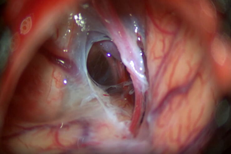
Optimal Visualization in Brain Surgery
This case study “Treatment of the Galassi type III arachnoid cyst with the M530 OHX surgical microscope from Leica Microsystems” documents the procedure step by step and shows the visualization…
Loading...

Spectroscopic Evaluation of Red Blood Cells
Hemoglobinopathies are a major healthcare problem. This study presents a possible diagnostic tool for thalassemia which is based on confocal spectroscopy. This approach exploits spectral detection and…
Loading...
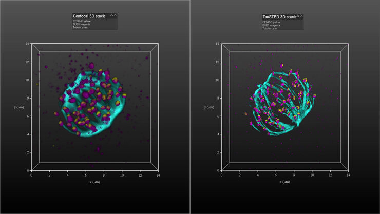
Kinetochore Assembly during Mitosis with TauSTED on 3D
Three-dimensional organization of the mitotic spindle together with the distribution of CENP-C and BUB1 based on TauSTED with multiple STED lines (592, 660 and 775 nm) can provide insights…
Loading...
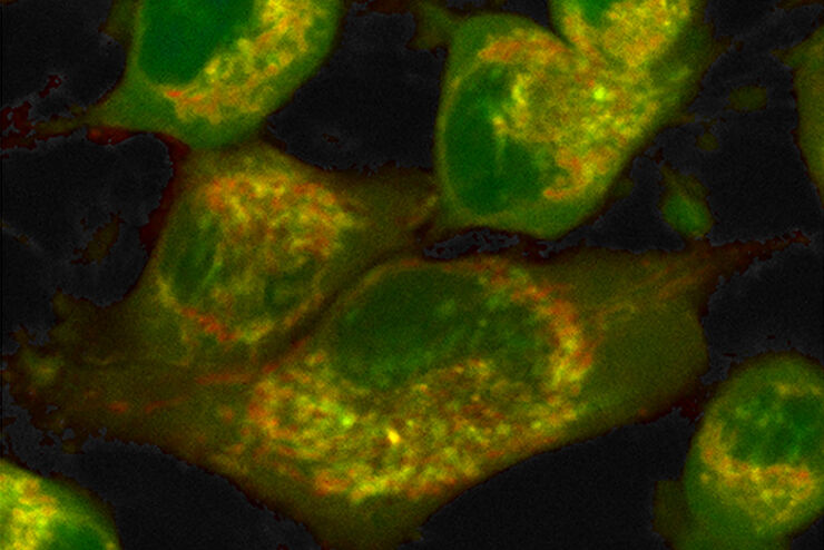
How to Quantify Changes in the Metabolic Status of Single Cells
Metabolic imaging based on fluorescence lifetime provides insights into the metabolic dynamics of cells, but its use has been limited as expertise in advanced microscopy techniques was needed.
Now,…
Loading...
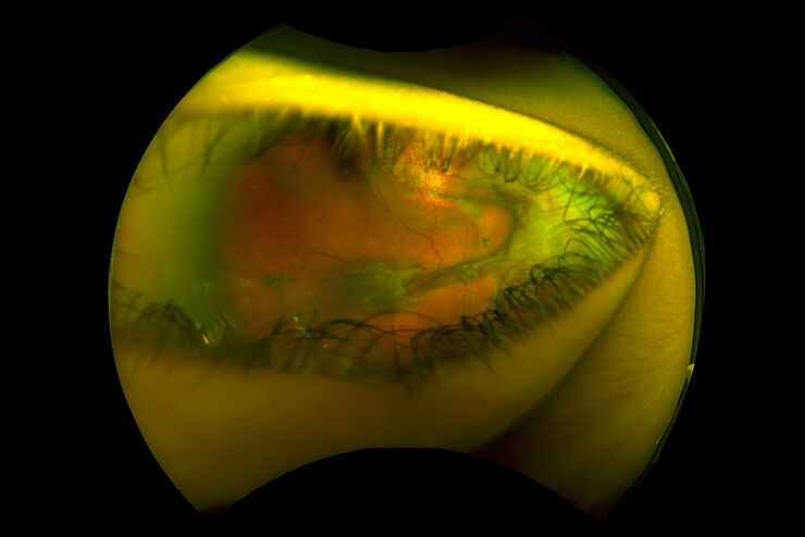
Intraoperative OCT-Assisted Surgical Management of Proliferative Vitreoretinopathy
Proliferative vitreoretinopathy (PVR) is a plague to patients and their surgeons after recent rhegmatogenous retinal detachment (RD). Despite excellent initial surgical outcomes, it is the most common…
Loading...
Investigating Synapses in Brain Slices with Enhanced Functional Electron Microscopy
A fundamental question of neuroscience is: what is the relationship between structural and functional properties of synapses? Over the last few decades, electrophysiology has shed light on synaptic…
Loading...

GLOW800 Augmented Reality Fluorescence in AVM (Arteriovenous Malformation) Treatment
In this case study Prof. Dr. Feres Chaddad talks about the treatment of AVMs. It illustrates how the Augmented Reality Fluorescence GLOW800 can help surgeons during microsurgical resection by…
Loading...
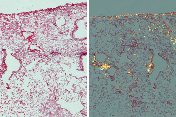
Studying Pulmonary Fibrosis
The results shown in this article demonstrate that fibrotic and non-fibrotic regions of collagen present in mouse lung tissue can be distinguished better with polarized light compared to brightfield.…
Loading...

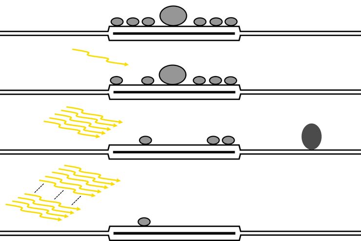
Keeping Particulate Contamination Under Control in Pharmaceutical Products
This article describes how a 2-methods-in-1 solution combining optical microscopy and laser induced breakdown spectroscopy (LIBS) can be utilized for identification of particulate contaminants in the…
