
Science Lab
Science Lab
Das Wissensportal von Leica Microsystems bietet Ihnen Wissens- und Lehrmaterial zu den Themen der Mikroskopie. Die Inhalte sind so konzipiert, dass sie Einsteiger, erfahrene Praktiker und Wissenschaftler gleichermaßen bei ihrem alltäglichen Vorgehen und Experimenten unterstützen. Entdecken Sie interaktive Tutorials und Anwendungsberichte, erfahren Sie mehr über die Grundlagen der Mikroskopie und High-End-Technologien - werden Sie Teil der Science Lab Community und teilen Sie Ihr Wissen!
Filter articles
Tags
Berichtstyp
Produkte
Loading...

Virologie
Liegt Ihr Forschungsschwerpunkt auf Virusinfektionen und -krankheiten? Erfahren Sie, wie Sie mit Lösungen für Bildgebung und Probenvorbereitung von Leica Microsystems mehr Erkenntnisse in der…
Loading...
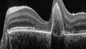
A Guide to OCT
Leica Optical Coherence Tomography (OCT) systems support ophthalmologists, ophthalmic surgeons, and researchers with easy-to-use, high-quality imaging technology.
Loading...

Zebrafisch-Forschung
Für optimale Ergebnisse während der Bewertung, Sortierung, Manipulation und Bildgebung von Modellorganismen ist es entscheidend feine Details und Strukturen genauestens zu erkennen. Das bildet die…
Loading...

Kryo-Elektronen-Tomographie
Mit der Kryo-Elektronentomographie (CryoET) lassen sich Biomoleküle in ihrer zellulären Umgebung mit einer noch nie dagewesenen Auflösung von weniger als einem Nanometer auflösen.
Loading...

A Guide to Spatial Biology
What is spatial biology, and how can researchers leverage its tools to meet the growing demands of biological questions in the post-omics era? This article provides a brief overview of spatial biology…
Loading...

Technical Terms for Digital Microscope Cameras and Image Analysis
Learn more about the basic principles behind digital microscope camera technologies, how digital cameras work, and take advantage of a reference list of technical terms from this article.
Loading...

Wichtige Faktoren, die Sie bei der Auswahl eines Stereomikroskops berücksichtigen sollten
Stereomikroskope zeichnen sich durch ihre Fähigkeit aus, einen 3D-Eindruck der Probe zu erzeugen. Daher eignen sie sich besonders gut für Inspektion und Nacharbeit, Qualitätskontrolle, Forschung und…
Loading...
 in 14 x 18 tiles. Lifetime gives an additional contrast that allows to differentiate different structures in histological stainings.")
Leitfaden zur Fluoreszenzlebensdauer-Imaging-Mikroskopie (FLIM)
Die Fluoreszenzlebensdauer ist ein Maß dafür, wie lange ein Fluorophor im Durchschnitt in seinem angeregten Zustand verbleibt, bevor er durch Aussendung eines Fluoreszenzphotons in den Grundzustand…
