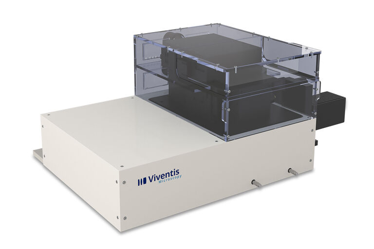
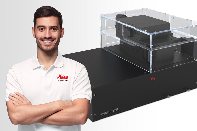
Viventis LS2 Live Microscope à fluorescence à feuillet de lumière à double détection
Le microscope à fluorescence à feuillet de lumière Viventis LS2 Live combine l’imagerie multivue et multiposition pour étudier les échantillons vivants.
Commencez votre parcours pour découvrir une imagerie profonde et à long terme qui révèle les détails et la dynamique complexes des systèmes biologiques.
Explorez la vie en profondeur
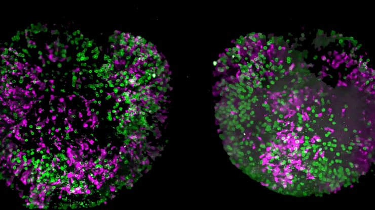
Le microscope Viventis LS2 Live vous aide à approfondir la compréhension de votre échantillon dans toute son épaisseur, grâce à sa résolution spatio-temporelle accrue.
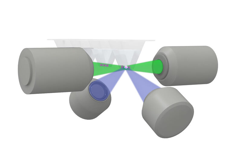
Atteignez une imagerie volumétrique détaillée et complète de l’échantillon grâce à une combinaison brevetée de
- Double illumination
- Double détection
- Multi-position
- Ouvrez le porte-échantillons supérieur
Vous pouvez même imager de grands échantillons diffusifs au cours du temps avec une qualité exceptionnelle pour une analyse post-acquisition significative, tout en minimisant la dose de lumière et en maintenant l’accès aux échantillons.
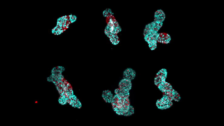
Etudiez plusieurs échantillons vivants en même temps
Obtenez plus de données à partir d’une seule expérience. Collectez les données de plusieurs échantillons en parallèle dans différentes conditions en un seul time-lapse. La configuration ouverte du microscope Viventis LS2 Live améliore votre imagerie par feuillet de lumière avec un débit plus élevé et des capacités d'acquisitions multipositions.
Elle permet de
- Monter facilement les échantillons
- Imager dans des conditions physiologiques avec seulement des modifications mineures du protocole
- Renouveler le milieu de culture, même pendant un time-lapse
Explorer des événements biologiques en imagerie à long terme
Dans une variété d'organismes modèles, y compris les organoïdes du cancer de l’intestin, du foie et du côlon humain et les embryons de poisson-zèbre, la technologie de feuillet de lumière du microscope Viventis LS2 Live fournit une qualité d’image élevée tout en préservant la viabilité de l’échantillon.
De plus, la solution d’incubation avancée et le remplacement facile du milieu de culture, même pendant une expérience en cours, préservent les conditions physiologiques. Gérez facilement des conditions expérimentales encore plus complexes. Pour étudier les réponses biologiques et garantir des résultats spécifiques, vous pouvez ajouter des traitements et utiliser un dispositif photomanipulation en option pendant les time-lapses
Adaptez la technologie du feuillet lumière à vos besoins
Le microscope Viventis LS2 Live vous aide à ajuster l’épaisseur du faisceau gaussien scanné (DLSM). Vous pouvez adapter le feuillet de lumière en mettant l’accent sur la résolution ou le champ de vision.
Son logiciel flexible vous permet de modifier les paramètres pendant les time-lapses. De plus, une interface de programmation d’application (API) Python vous permet de coder et d'utiliser des macros personnalisées pour vos propres idées expérimentales.
Pour empêcher votre échantillon de sortir du champ de vision (FOV) pendantun time-lapse, le tracking intelligent des objets modifie les coordonnées de la platine.
Un soutien complet de votre workflow au service de votre réussite
Nous assurons la bonne continuité de vos travaux de Recherche dans le monde entier grâce à un service après-vente de premier ordre entièrement dédié à la microscopie depuis plus de 175 ans.
Caractéristiques principales
- Équipe Leica : + de 500 experts en service après-vente et applications
- Formation Leica : programme de certification d’usine à 4 niveaux
- Logistique Leica : 5 centres régionaux pour les pièces d’origine
- Leica OneCall : Assistance téléphonique de niveau PhD
