
Science Lab
Science Lab
Bienvenue sur le portail de connaissances de Leica Microsystems. Vous y trouverez des recherches scientifiques et du matériel didactique sur le thème de la microscopie. Le portail aide les débutants, les praticiens expérimentés et les scientifiques dans leur travail quotidien et leurs expériences. Explorez les didacticiels interactifs et les notes d'application, découvrez les bases de la microscopie ainsi que les technologies de pointe. Faites partie de la communauté Science Lab et partagez votre expertise.
Filter articles
Tags
Type de publication
Produits
Loading...

Improving Zebrafish-Embryo Screening with Fast, High-Contrast Imaging
Discover from this article how screening of transgenic zebrafish embryos is boosted with high-speed, high-contrast imaging using the DM6 B microscope, ensuring accurate targeting for developmental…
Loading...

Epi-Illumination Fluorescence and Reflection-Contrast Microscopy
This article discusses the development of epi-illumination and reflection contrast for fluorescence microscopy concerning life-science applications. Much was done by the Ploem research group…
Loading...

Differential Interference Contrast (DIC) Microscopy
This article demonstrates how differential interference contrast (DIC) can be actually better than brightfield illumination when using microscopy to image unstained biological specimens.
Loading...
 cells taken with phase contrast.")
Phase Contrast and Microscopy
This article explains phase contrast, an optical microscopy technique, which reveals fine details of unstained, transparent specimens that are difficult to see with common brightfield illumination.
Loading...
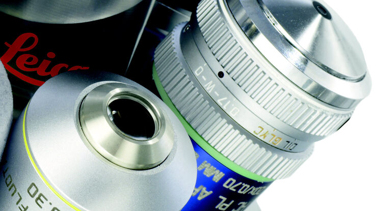
Immersion Objectives
How an immersion objective, which has a liquid medium between it and the specimen being observed, helps increase the numerical aperture and microscope resolution is explained in this article.
Loading...

A Guide to Darkfield Microscopes
A darkfield microscope offers a way to view the structures of many types of biological specimens in greater contrast without the need of stains.
Loading...
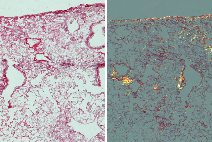
Studying Pulmonary Fibrosis
The results shown in this article demonstrate that fibrotic and non-fibrotic regions of collagen present in mouse lung tissue can be distinguished better with polarized light compared to brightfield.…
Loading...
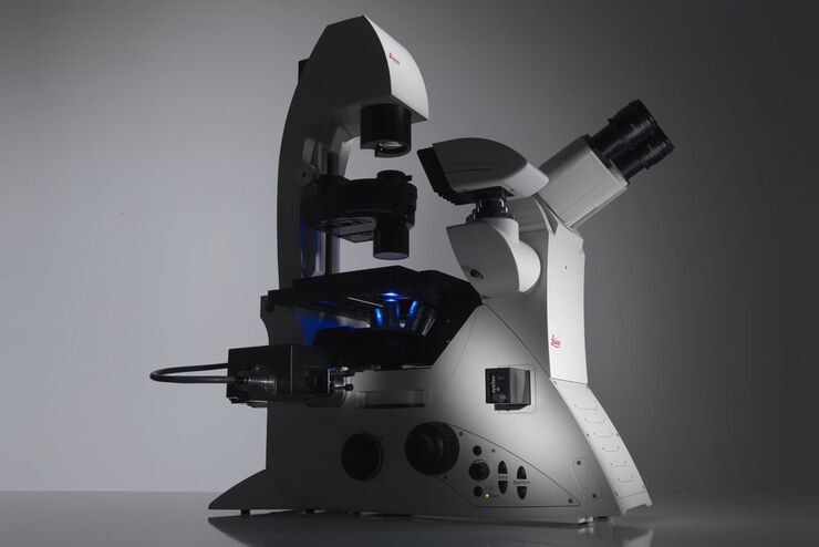
Factors to Consider When Selecting a Research Microscope
An optical microscope is often one of the central devices in a life-science research lab. It can be used for various applications which shed light on many scientific questions. Thereby the…
Loading...

Virologie
Vos recherches portent sur les infections et les maladies virales ? Découvrez comment renforcer vos connaissances en virologie grâce aux solutions d’imagerie et de préparation d’échantillons de Leica…
