TCS SP5 II 共焦点顕微鏡<br>
Multi-color imaging in whole mount specimen
Platyneris spec.
blue: nuclei, DAPI; green: Actin/muscles, TRITC; red: Tubulin, Alexa 633
Courtesy of: Dr. Evgeny Tsitrin, Institute of Developmental Biology RAS, Moscow, Russia
Imaging and manipulation with new specific optics
The optics of the Leica TCS SP5 II is optimized for imaging and manipulation. The beam expander allows for switching between outstanding precision and high bleach power. This means that there is no impairment of optical properties if bleaching or imaging.
High resolution and high speed confocal imaging
The Leica Tandem Scanner combines two technological solutions in one system: a conventional scanning system - ideal for morphological imaging and three dimensional structure analyses, and a resonant scanning system - the best solution for high speed confocal imaging.Merging both systems into one…
The fastest true confocal
The combined efficiency of the Leica SP® detection system and the Leica AOBS® guarantees best signal to noise – which means clear and crisp images and least bleaching during image acquisition. Easy operation and a minimum of maintenance save time and money and ensures more results in less time.
Electrophysiology with Leica DM6000 CFS
Small neuron network: rat brain slice, layer 5. Loading of dyes by single cell electroporation.
Loading of dyes by single cell electroporationred: Interneurons Alexa 594; green: Pyramidal Cell Oregon Bapta 1 (calcium sensitive)
Z = 123 µm; two-photon excitation; detection with a 2 channel NDD
Courte…
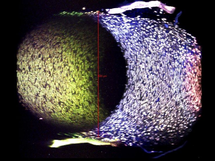
Deep tissue imaging with multi-photon microscopy
Artery of the mouse: Excitation at 839 nm, 3-channel acquisition: autofluorescence of elastin (blue), Syto13 for nuclei of cells in the vascular wall (green/white), eosin auto-fluorescence (red); zoom 1. Imaging depth 650 µm. Preparation: Common cartoid arteries from mice are carefully dissected,…


