
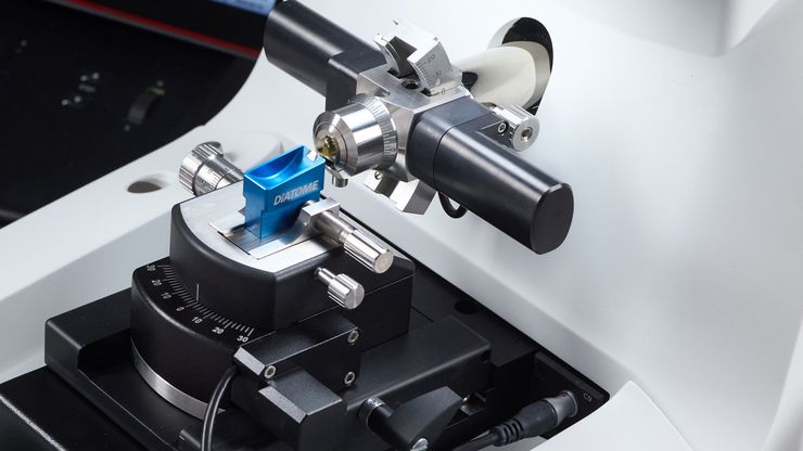
Mastering Polymer Sectioning with Helmut Gnaegi
When it comes to ultramicrotomy, few names carry the weight of Helmut Gnaegi. As co-founder of Diatome, a global leader in diamond knife technology, Helmut has spent decades refining the art and…

Mica: A Game-changer for Collaborative Research at Imperial College London
This interview highlights the transformative impact of Mica at Imperial College London. Scientists explain how Mica has been a game-changer, expanding research possibilities and facilitating…

Exploring Microbial Worlds: Spatial Interactions in 3D Food Matrices
The Micalis Institute is a joint research unit in collaboration with INRAE, AgroParisTech, and Université Paris-Saclay. Its mission is to develop innovative research in the field of food microbiology…

Advancing Uterine Regenerative Therapies with Endometrial Organoids
Prof. Kang's group investigates important factors that determine the uterine microenvironment in which embryo insertion and pregnancy are successfully maintained. They are working to develop new…

How did Laser Microdissection enable Pioneering Neuroscience Research?
Dr. Marta Paterlini, a Senior Scientist at the Karolinska Institute, shares her experience of using laser microdissection (LMD) in groundbreaking research into adult human neurogenesis and offers…

Multiplexing with Luke Gammon: Advance your Spatial Biology Research
Learn how multiplexing imaging and spatial biology can help researchers better understand complex biological systems. In this interview, Dr. Gammon and Dr. Pointu of Leica Microsystems discuss pain…

Examining Developmental Processes In Cancer Organoids
Interview: Prof. Bausch and Dr. Pastucha, Technical University of Munich, discuss using microscopy to study development of organoids, stem cells, and other relevant disease models for biomedical…
. Image courtesy of Prof. Hui Guo, School of Life Sciences, Central South University, China")
機械受容性経路とシナプス経路の研究に顕微鏡がいかに役立つか
このポッドキャストでは、Tobi Langenhan教授は、顕微鏡を使ってシナプスのタンパク質集合体を調べるなど、接着型GPCRの機械受容特性の研究を通して、タンパク質のダイナミクスとその空間的相互作用に精通されています。 Abdullah…

Applying AI and Machine Learning in Microscopy and Image Analysis
Prof. Emma Lundberg is a professor in cell biology proteomics at KTH Royal Institute of Technology, Sweden. She is also the director of the Cell Atlas, an integral part of the Swedish-based Human…
