STELLARIS DIVE
공초점 레이저 현미경
제품소개
홈
Leica Microsystems
STELLARIS DIVE 다광자 현미경
다색 다광자로 진실에 더 가까이
최신 기사를 읽어 보세요
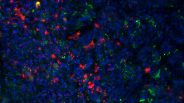
Intravital Microscopy of Cancer
Join our guest speaker Prof Dr Jacco van Rheenen, as he presents his work on the identity, behavior and fate of cells that drive the initiation and progression of cancer.
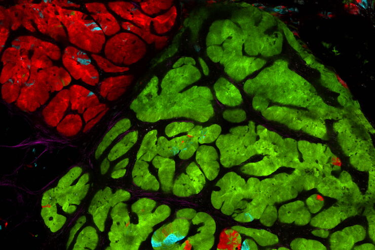
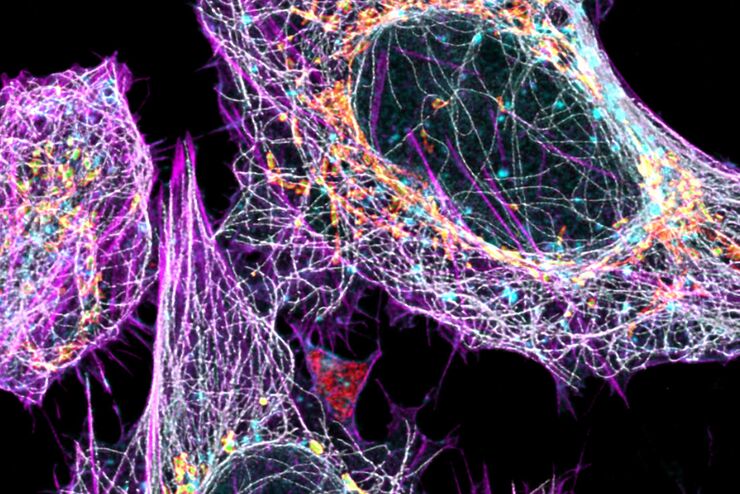
Multicolor Image Gallery
Fluorescence multicolor microscopy, which is one aspect of multiplex imaging, allows for the observation and analysis of multiple elements within the same sample – each tagged with a different…
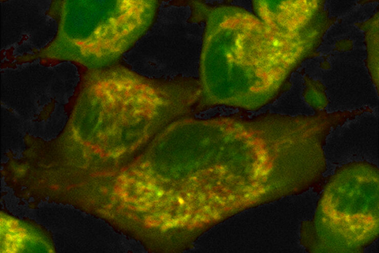
How to Quantify Changes in the Metabolic Status of Single Cells
Metabolic imaging based on fluorescence lifetime provides insights into the metabolic dynamics of cells, but its use has been limited as expertise in advanced microscopy techniques was needed.
Now,…
연구 분야의 모델 유기체
모델 유기체는 연구자들이 특정한 생물학적 과정을 연구하기 위해 사용하는 종입니다. 이들은 인간과 유사한 유전적 특성을 가지고 있으며, 유전학, 발달생물학, 신경과학 같은 연구 분야에서 일반적으로 사용됩니다. 유기체 모델은 일반적으로 실험실 환경에서 쉬운 유지와 번식, 짧은 세대 주기 또는 특정 형질이나 질병을 연구하기 위한 돌연변이 생성 능력 때문에…
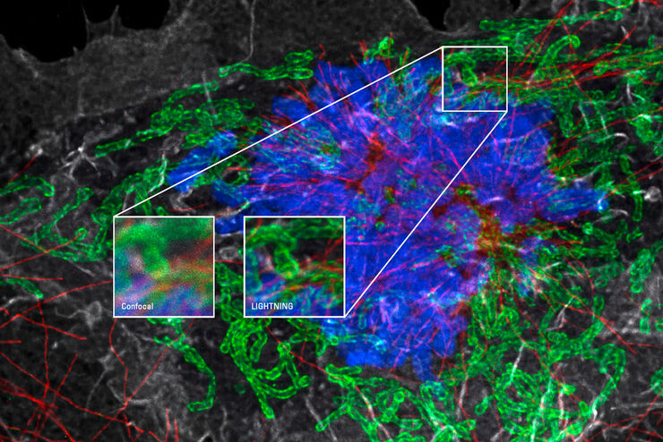
LIGHTNING으로 시료에서 최대한의 정보를 얻으세요
LIGHTNING은 숨겨진 정보를 추출하는 조절 가능한 프로세스를 사용하여 미세한 구조와 세부 정보도 완전히 자동으로 표현해 냅니다. 전체 이미지에 포괄적인 파라미터 집합을 사용하는 기존 기술과 달리, LIGHTNING은 각 복셀에 적합한 파라미터 집합만을 계산하여 최고의 정확도로 모든 세부 정보를 파악합니다.
바이러스학
연구의 관심 분야가 바이러스 감염과 질병에 집중되어 있습니까? 라이카마이크로시스템즈의 이미징 및 샘플 준비 솔루션을 통해 바이러스학에 관한 통찰력을 얻는 방법을 알아보세요.
Principles of Multiphoton Microscopy for Deep Tissue Imaging
This tutorial explains the principles of multiphoton microscopy for deep tissue imaging. Multiphoton microscopy uses excitation wavelengths in the infrared taking advantage of the reduced scattering…
New Standard in Electrophysiology and Deep Tissue Imaging
The function of nerve and muscle cells relies on ionic currents flowing through ion channels. These ion channels play a major role in cell physiology. One way to investigate ion channels is to use…
Fields of Application
오가노이드와 3D 세포 배양
최근 생명과학 연구에서 가장 흥미로운 발전 중 하나는 오가노이드, 스페로이드 또는 장기 칩 모델과 같은 3D 세포 배양 시스템의 개발입니다. 3D 세포 배양이란 세포가 3차원에서 성장하고 주변 환경과 상호작용할 수 있는 인위적인 환경입니다. 이러한 조건은 체내 상태와 유사합니다.
