DM IL LED
도립
광학 현미경
제품소개
홈
Leica Microsystems
DM IL LED Inverted Laboratory Microscope
최신 기사를 읽어 보세요
Microscopy and AI Solutions for 2D Cell Culture
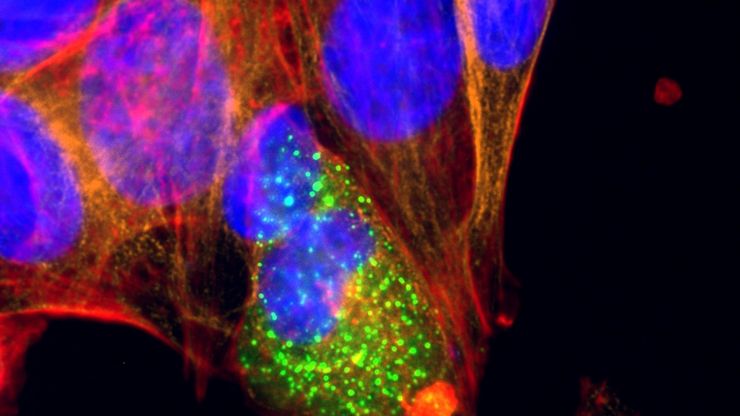
This eBook explores the integration of microscopy and AI technologies in 2D cell culture workflows. It highlights how traditional imaging methods—such as brightfield, phase contrast, and…
Phase Contrast and Microscopy
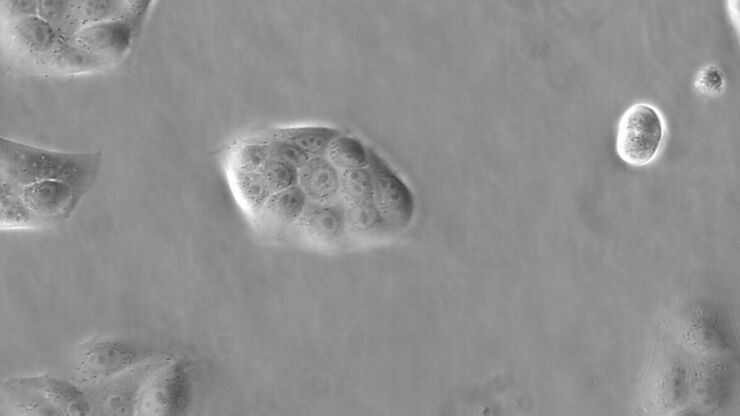
This article explains phase contrast, an optical microscopy technique, which reveals fine details of unstained, transparent specimens that are difficult to see with common brightfield illumination.
How to do a Proper Cell Culture Quick Check
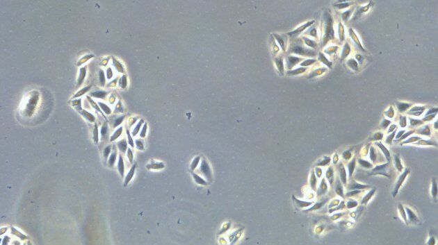
In order to successfully work with mammalian cell lines, they must be grown under controlled conditions and require their own specific growth medium. In addition, to guarantee consistency their growth…
Microscopy in Virology
The coronavirus SARS-CoV-2, causing the Covid-19 disease effects our world in all aspects. Research to find immunization and treatment methods, in other words to fight this virus, gained highest…
Introduction to Mammalian Cell Culture
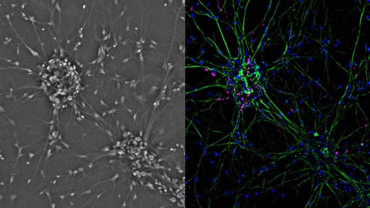
Mammalian cell culture is one of the basic pillars of life sciences. Without the ability to grow cells in the lab, the fast progress in disciplines like cell biology, immunology, or cancer research…
Introduction to Widefield Microscopy
This article gives an introduction to widefield microscopy, one of the most basic and commonly used microscopy techniques. It also shows the basic differences between widefield and confocal…
Fields of Application
세포 배양
실험실 조건에서의 세포 배양은 세포생물학, 암 연구, 발생생물학을 비롯한 모든 종류의 생명 과학 및 제약 연구 분야에 속한 과학자들을 위한 기초입니다. Leica가 실험실에서 동물 세포를 배양하는 데 어떤 도움이 될 수 있는지 알아보십시오.
