Infinity TIRF
부속품
제품소개
홈
Leica Microsystems
Infinity TIRF DMi8 S 모듈
숨겨진 것까지 관찰
최신 기사를 읽어 보세요
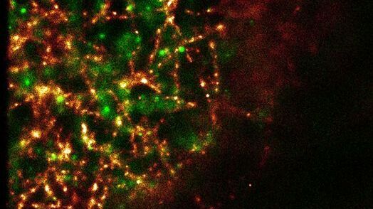
, and trafficking vesicles labeled with CF594 (cyan - Biotium).")
A Guide to Super-Resolution
Find out more about Leica super-resolution microscopy solutions and how they can empower you to visualize in fine detail subcellular structures and dynamics.

바이러스학
연구의 관심 분야가 바이러스 감염과 질병에 집중되어 있습니까? 라이카마이크로시스템즈의 이미징 및 샘플 준비 솔루션을 통해 바이러스학에 관한 통찰력을 얻는 방법을 알아보세요.

Universal PAINT – Dynamic Super-Resolution Microscopy
Super-resolution microscopy techniques have revolutionized biology for the last ten years. With their help cellular components can now be visualized at the size of a protein. Nevertheless, imaging…
Sample Preparation for GSDIM Localization Microscopy – Protocols and Tips
The widefield super-resolution technique GSDIM (Ground State Depletion followed by individual molecule return) is a localization microscopy technique that is capable of resolving details as small as…
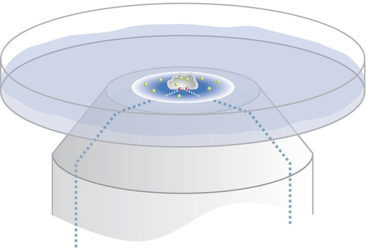
Controlling the TIRF Penetration Depth is Mandatory for Reproducible Results
The main feature of total internal reflection fluorescence (TIRF) microscopy is the employment of an evanescent wave for the excitation of fluorophores instead of using direct light. A property of the…

Total Internal Reflection Fluorescence (TIRF) Microscopy
Total internal reflection fluorescence (TIRF) is a special technique in fluorescence microscopy developed by Daniel Axelrod at the University of Michigan, Ann Arbor in the early 1980s. TIRF microscopy…
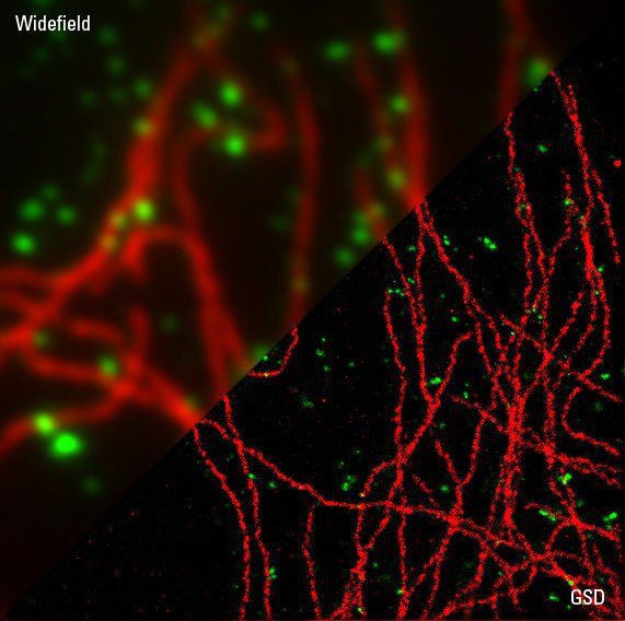
Super-Resolution GSDIM Microscopy
The nanoscopic technique GSDIM (ground state depletion microscopy followed by individual molecule return) provides a detailed image of the spatial arrangement of proteins and other biomolecules within…
적용 분야
세포생물학
인간의 건강과 질병을 기준으로 세포를 이해하는 것에 연구의 초점이 맞추어져 있다면 관심 세포를 시공간 및 분자 측면에서 자세히 조사하는 것은 매우 중요합니다. 이는 현미경이 세포생물학에서 매우 중요한 도구인 이유입니다. 현미경을 사용하면 세포 기관과 고분자를 분석할 뿐만 아니라, 시료의 구조적 환경 내에서 시료를 자세히 연구할 수 있습니다. 세포생물학…
