LAS X Life Science
현미경 소프트웨어
제품소개
홈
Leica Microsystems
LAS X Life Science 생명과학용 소프트웨어 플랫폼
모두를 위한 하나
최신 기사를 읽어 보세요
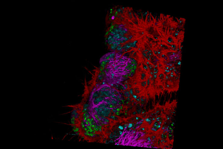
Find Relevant Specimen Details from Overviews
Switch from searching image by image to seeing the full overview of samples quickly and identifying the important specimen details instantly with confocal microscopy. Use that knowledge to set up…
Artificial Intelligence and Confocal Microscopy – What You Need to Know
This list of frequently asked questions provides “hands-on” answers and is a supplement to the introductory article about Dynamic Signal Enhancement powered by Aivia "How Artificial Intelligence…
How Artificial Intelligence Enhances Confocal Imaging
In this article, we show how artificial intelligence (AI) can enhance your imaging experiments. Namely, how Dynamic Signal Enhancement powered by Aivia improves image quality while capturing the…
Fluorescence Lifetime-based Imaging Gallery
Confocal microscopy relies on the effective excitation of fluorescence probes and the efficient collection of photons emitted from the fluorescence process. One aspect of fluorescence is the emission…
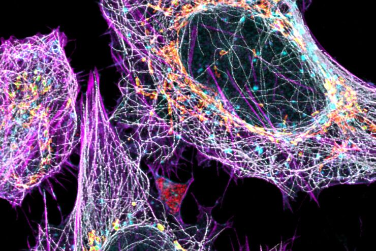
Multicolor Image Gallery
Fluorescence multicolor microscopy, which is one aspect of multiplex imaging, allows for the observation and analysis of multiple elements within the same sample – each tagged with a different…
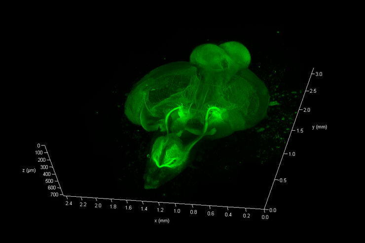
Zebrafish Brain - Whole Organ Imaging at High Resolution
Structural information is key when one seeks to understand complex biological systems, and one of the most complex biological structures is the vertebrate central nervous system. To image a complete…
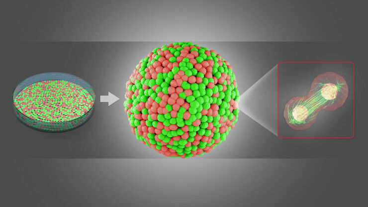
Improve 3D Cell Biology Workflow with Light Sheet Microscopy
Understanding the sub-cellular mechanisms in carcinogenesis is of crucial importance for cancer treatment. Popular cellular models comprise cancer cells grown as monolayers. But this approach…
적용 분야
오가노이드와 3D 세포 배양
최근 생명과학 연구에서 가장 흥미로운 발전 중 하나는 오가노이드, 스페로이드 또는 장기 칩 모델과 같은 3D 세포 배양 시스템의 개발입니다. 3D 세포 배양이란 세포가 3차원에서 성장하고 주변 환경과 상호작용할 수 있는 인위적인 환경입니다. 이러한 조건은 체내 상태와 유사합니다.


