K3C & K3M
현미경 카메라
제품소개
홈
Leica Microsystems
K3C & K3M 현미경 카메라 시리즈
생명과학 및 산업 이미징 애플리케이션 및 분석용
최신 기사를 읽어 보세요
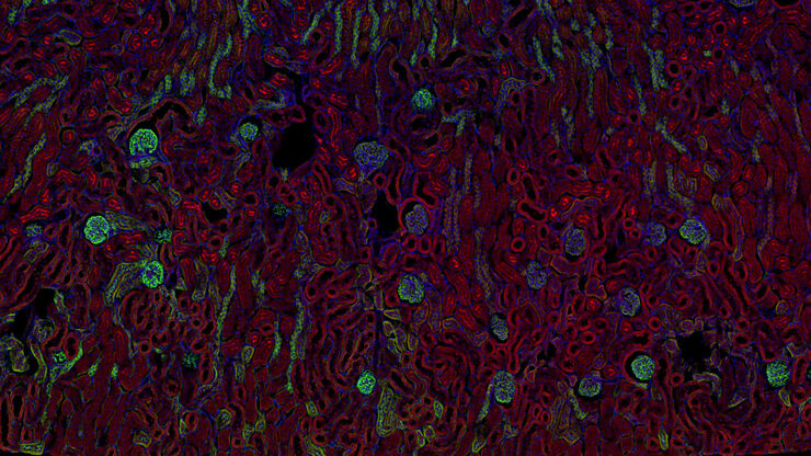
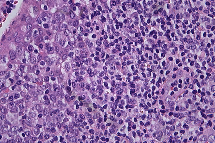
암 연구
암은 성장 통제에 결함이 있는 세포에 의해 발생하는 복잡하고 이질적인 질병입니다. 하나 또는 한 그룹의 세포에서 일어나는 유전적 또는 후생적 변화가 정상적인 기능을 방해하고, 자율적이고 통제되지 않는 세포 성장과 증식을 초래합니다
Technical Terms for Digital Microscope Cameras and Image Analysis
Learn more about the basic principles behind digital microscope camera technologies, how digital cameras work, and take advantage of a reference list of technical terms from this article.
Understanding Clearly the Magnification of Microscopy
To help users better understand the magnification of microscopy and how to determine the useful range of magnification values for digital microscopes, this article provides helpful guidelines.
Life Science Research: Which Microscope Camera is Right for You?
Deciding which microscope camera best fits your experimental needs can be daunting. This guide presents the key factors to consider when selecting the right camera for your life science research.
Factors to Consider when Selecting Clinical Microscopes
What matters if you would like to purchase a clinical microscope? Learn how to arrive at the best buying decision from our Science Lab Article.
Clinical Microscopy: Considerations on Camera Selection
The need for images in pathology laboratories has significantly increased over the past few years, be it in histopathology, cytology, hematology, clinical microbiology, or other applications. They…
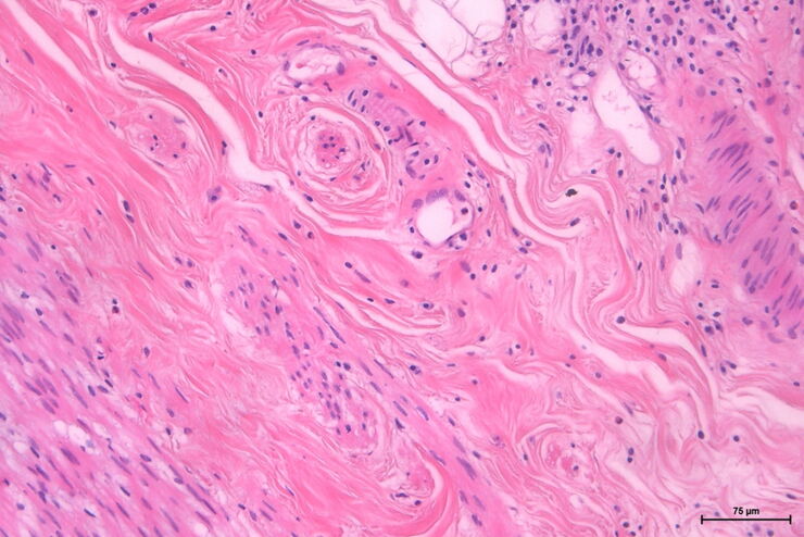
The Time to Diagnosis is Crucial in Clinical Pathology
Abnormalities in tissues and fluids - that’s what pathologists are looking for when they examine specimens under the microscope. What they see and deduce from their findings is highly influential, as…
적용 분야
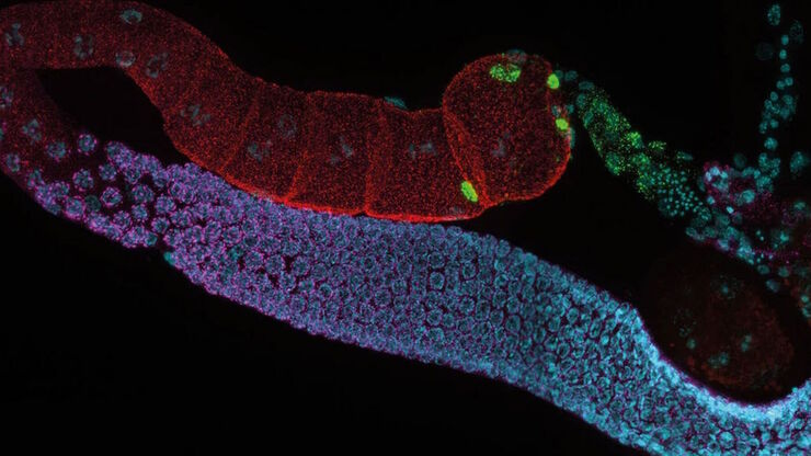
생명 과학 연구
라이카사의 생명과학사업부는 미세구조의 시각화 및 분석을 위한 혁신 기술 및 기술 전문성을 원하는 scientific community 를 충족시키는 imaging을 지원할 수있습니다. 라이키의 고객을 과학분야의 선두자로 이끌어내는것에 관심이 있습니다.
산업용 현미경 시장
가동 시간을 최대화하고 목표를 달성하면 수익에 효율적으로 도움이 됩니다. Leica 현미경 솔루션은 가장 작은 샘플 세부 사항에 대한 통찰력을 제공할 뿐만 아니라 결과를 빠르고 안정적으로 분석, 문서화 및 보고할 수 있습니다. Leica Microsystems는 다양한 애플리케이션 요구 사항을 충족하는 데 도움이 되는 광범위한 솔루션과 전문가 지원을…
