K5C
현미경 카메라
제품소개
홈
Leica Microsystems
K5C 컬러 CMOS 카메라
강력하고 집중적인 연구 선택
최신 기사를 읽어 보세요
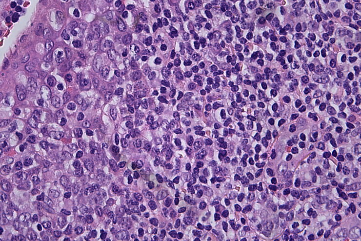
Clinical Microscopy: Considerations on Camera Selection
The need for images in pathology laboratories has significantly increased over the past few years, be it in histopathology, cytology, hematology, clinical microbiology, or other applications. They…
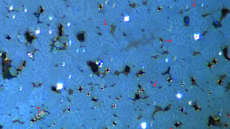
Rapidly Visualizing Magnetic Domains in Steel with Kerr Microscopy
The rotation of polarized light after interaction with magnetic domains in a material, known as the Kerr effect, enables the investigation of magnetized samples with Kerr microscopy. It allows rapid…
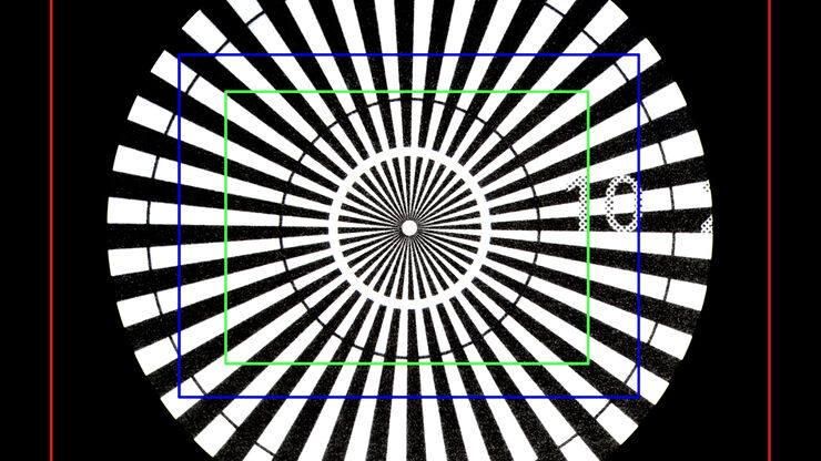
Understanding Clearly the Magnification of Microscopy
To help users better understand the magnification of microscopy and how to determine the useful range of magnification values for digital microscopes, this article provides helpful guidelines.
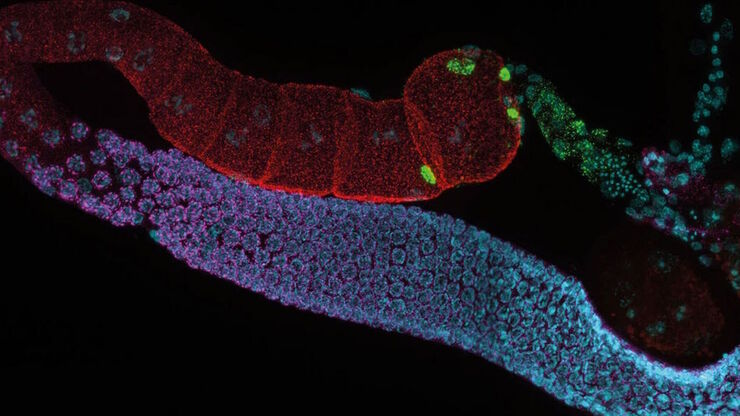
Life Science Research: Which Microscope Camera is Right for You?
Deciding which microscope camera best fits your experimental needs can be daunting. This guide presents the key factors to consider when selecting the right camera for your life science research.
Top Issues Related to Standards for Rating Non-Metallic Inclusions in Steel
Supplying components and products made of steel to users worldwide can require that a single batch be compliant with multiple steel quality standards. This user demand creates significant challenges…
Fields of Application
생명 과학 연구
라이카사의 생명과학사업부는 미세구조의 시각화 및 분석을 위한 혁신 기술 및 기술 전문성을 원하는 scientific community 를 충족시키는 imaging을 지원할 수있습니다. 라이키의 고객을 과학분야의 선두자로 이끌어내는것에 관심이 있습니다.


