Viventis Deep
광시트 현미경
제품소개
홈
Leica Microsystems
Viventis Deep 듀얼 뷰 광시트 형광 현미경
살아있는 샘플을 전체 맥락으로 보기
최신 기사를 읽어 보세요
How to Study Gene Regulatory Networks in Embryonic Development
Join Dr. Andrea Boni by attending this on-demand webinar to explore how light-sheet microscopy revolutionizes developmental biology. This advanced imaging technique allows for high-speed, volumetric…
Dual-View LightSheet Microscope for Large Multicellular Systems
Visualizing the dynamics of complex multicellular systems is a fundamental goal in biology. To address the challenges of live imaging over large spatiotemporal scales, Franziska Moos et. al. present…
Download The Guide to Live Cell Imaging
In life science research, live cell imaging is an indispensable tool to visualize cells in a state as in vivo as possible. This E-book reviews a wide range of important considerations to take to…
연구 분야의 모델 유기체
모델 유기체는 연구자들이 특정한 생물학적 과정을 연구하기 위해 사용하는 종입니다. 이들은 인간과 유사한 유전적 특성을 가지고 있으며, 유전학, 발달생물학, 신경과학 같은 연구 분야에서 일반적으로 사용됩니다. 유기체 모델은 일반적으로 실험실 환경에서 쉬운 유지와 번식, 짧은 세대 주기 또는 특정 형질이나 질병을 연구하기 위한 돌연변이 생성 능력 때문에…
적용 분야
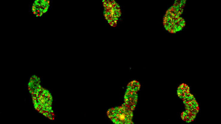
오가노이드와 3D 세포 배양
최근 생명과학 연구에서 가장 흥미로운 발전 중 하나는 오가노이드, 스페로이드 또는 장기 칩 모델과 같은 3D 세포 배양 시스템의 개발입니다. 3D 세포 배양이란 세포가 3차원에서 성장하고 주변 환경과 상호작용할 수 있는 인위적인 환경입니다. 이러한 조건은 체내 상태와 유사합니다.


