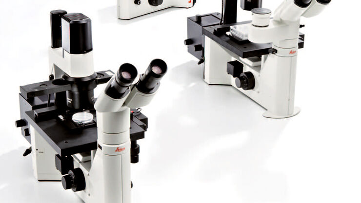
당사의 전문가들이 고객의 요구에 맞는 세포 배양 솔루션을 찾을 수 있도록 도와드립니다.
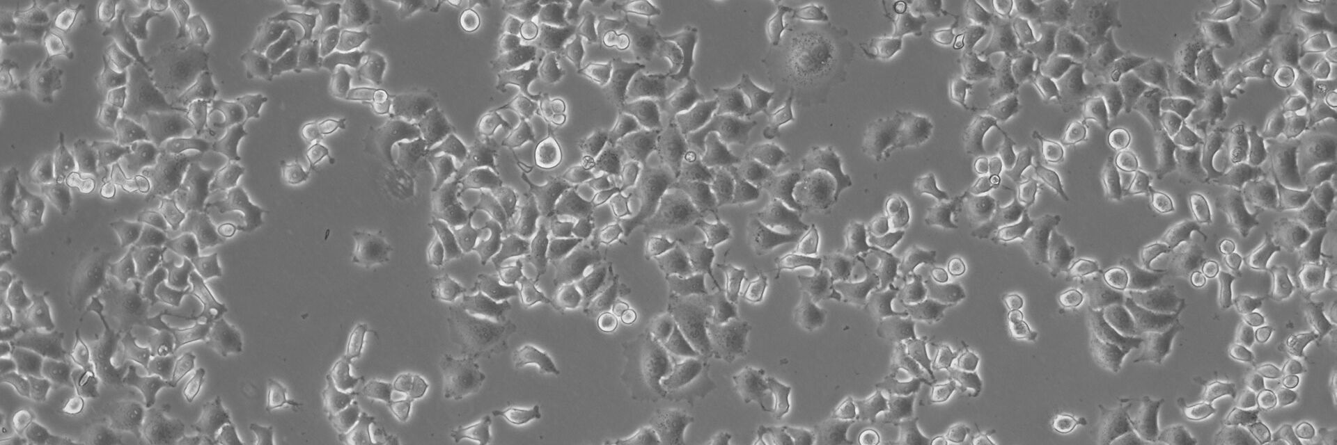
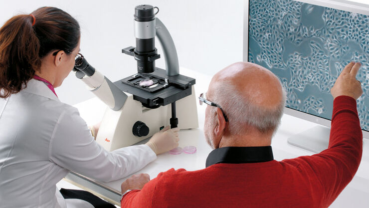
동물 세포는 본질적으로 콘트라스트가 매우 낮기 때문에 세포 배양 현미경은 위상차 등의 콘트라스트 방법을 지원해야 합니다. 여기에서 DIC(Differential Interference Contrast, 미분 간섭)는 세포 배양에 사용되는 플라스틱 용기와 함께 사용할 수 없기 때문에 도움이 되지 않습니다. DIC의 매우 좋은 대안은 플라스틱 용기와 함께 사용할 수 있고 특수한 대물렌즈나 프리즘이 필요하지 않은 IMC(Integrated Modulation Contrast, 통합 모듈레이션 콘트라스트)입니다. 뿐만 아니라, 세포 배양 현미경은 시간 손실을 막기 위해 취급이 쉽고 간편해야 합니다.
Leica 세포 배양 현미경은 사용이 쉽고 간편할 뿐만 아니라 각각의 요건에 부합하는 다양한 콘트라스트 방법을 제공합니다.
{{ question.questionText }}
답을 선택해 주세요!
베스트 매치
{{ resultProduct.header }}
{{ resultProduct.subheader }}
{{ resultProduct.description }}
{{ resultProduct.features }}
정보 패키지 요청하기
세포 배양 제품
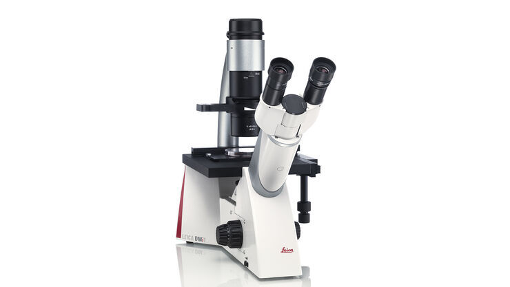
Inverted Microscope for Cell Culture Leica DMi1
Leica DMi1 도립현미경은 사용자의 특정한 작업 방식을 도와드립니다. 직관적인 조작방식은 사용자가 오로지 작업에만 집중할 수 있도록 편안함을 제공합니다. 사용자 원하는 기능을 선택하십시오! 필요에 따라 꼭 필요한 액세서리를 손쉽게 추가하실 수도 있습니다.
Inverted Laboratory Microscope Leica DM IL LED
Leica DM IL LED는 원하는 방식으로 시료를 모니터링하는 다양한 컨트라스트 방법을 제공합니다. 고품질 위상차, 뛰어난 모듈레이션 콘트라스트 및 선명한 형광 분석을 간편하게 이용할 수 있습니다. 견고한 안정성, 도구를 사용해 작업할 수 있는 넉넉한 공간, 대형 배양 플라스크를 수용할 수 있는 충분한 작업 거리, 뜨겁지 않은 안정적인 조명을 통해 현미경을 쉽고 편리하게 사용할 수 있습니다.
| 명시야 | 위상차 | DIC | IMC | 형광 | 배율 | 작업 거리 | 카메라 |
Leica DM IL LED | + | + | - | + | + | PH: 5x ~ 63x IMC: 10x, 20x, 32x, 40x | 40 mm, 80 mm | + (자유 선택) |
Leica DMi1 | + | + | - | - | - | 10x, 20x, 40x | 40 mm, 50 mm, 80 mm | + (통합) |
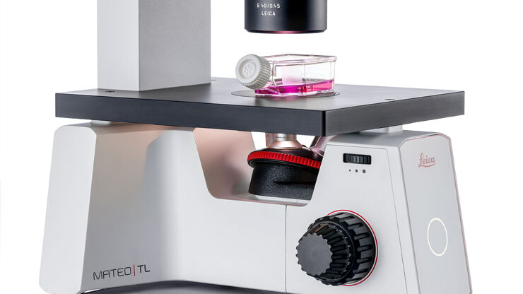
Mateo TL | + | + | - | - | - | 4x, 10x, 20x, 40x | 50 mm | + (통합) |
Mateo FL | + | + | - | - | + | 2.5x, 4x, 5x, 10x, 20x, 40x, 63x | 50 mm | + 듀얼 카메라 (통합) |
세포 배양 실험실 작업 전용 현미경
세포의 모양
실험실에서 배양한 동물 세포는 여러 기준으로 구분할 수 있습니다.
이러한 세포의 형태는 현미경으로 쉽게 식별이 가능합니다. Fibroblast-like cell은 양극성 또는 다극성이고 모양이 가늘고 긴 반면, Epithelial-like cell은 윤곽이 다각형입니다. 위의 두 세포와 반대로, Lymphoblast-like cell은 표면에 부착되지 않고 부유 상태로 배양됩니다.
세포 유형은 불멸화 세포, 초대배양 세포 그리고 줄기 세포로 세분화됩니다.
세포 조직은 단순한 2D 단일 배양에서 2D 공동 배양과 3D 스페로이드와 오가노이드에 이르기까지 다양합니다.
현미경 – 고급 요구사항
어떤 툴이 필요한가?
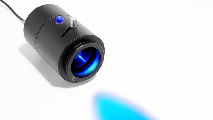
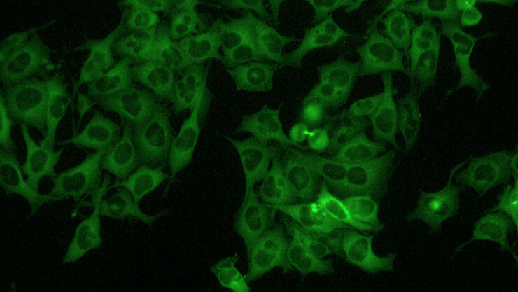
매우 일반적인 생물학적 접근법은 연구용 현미경으로 후속 검사를 하기 위해 형광 마커를 이용해 세포에 형질주입을 하는 것입니다. 형광 단백질을 다룰 경우에도 형질주입 효율 등을 제어하기 위해 세포 배양 현미경에 형광 옵션이 필요합니다.
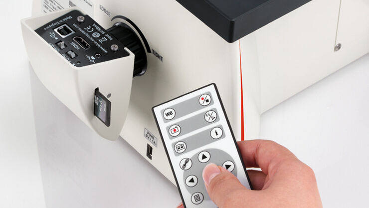
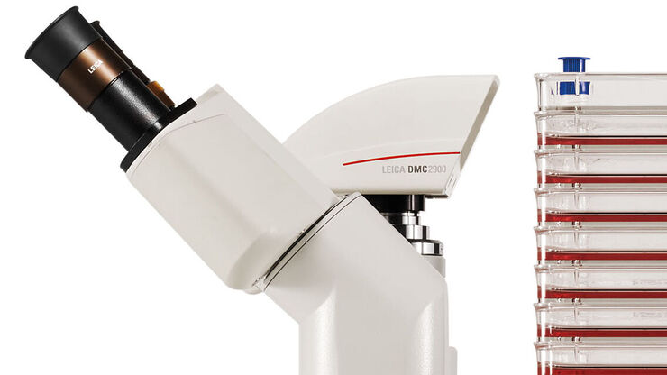
의미 있는 문서화와 표준화를 위해서는 현미경에 디지털 카메라가 있어야 하고 이상적으로는 인식한 데이터를 기록하고 분석할 수 있어야 합니다.
세포 배양 실험실에서는 공간이 문제이기 때문에 세포 배양 현미경이 너무 크면 안 됩니다(예: 후드 아래에 들어가야 함). 뿐만 아니라, 인큐베이터 안에서도 사용할 정도로 작고견고한 현미경이 최근의 추세입니다.
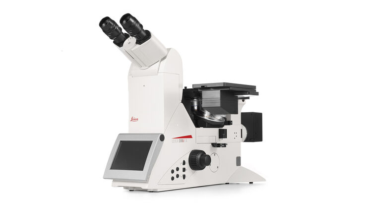
디쉬에서 단일 세포의 발달을 정밀하게 관찰하든, 여러 분석법을 통해 선별하든, 단일 분자 해상도를 얻든, 복잡한 프로세스의 작용을 파악하든 상관없이, DMi8 S 시스템을 사용하면 더 많은 것을 더 빠르게 숨겨진 것까지 관찰할 수 있습니다
