신경과학 연구

신경과학 분야에서는 종종 신경계를 더 잘 이해하기 위해 현미경을 사용하여 까다롭고 복잡한 시료를 조사해야 합니다.

 Neuroscience-Solutions-Res-.jpg

신경과학 분야에서는 종종 신경계를 더 잘 이해하기 위해 현미경을 사용하여 까다롭고 복잡한 시료를 조사해야 합니다. 라이카마이크로시스템즈는 이러한 문제를 극복할 수 있는 포괄적인 이미지 솔루션을 제공합니다.

신경과학 연구

신경과학은 신경계의 구조와 기능에 대한 연구를 포함하는 종합적인 분야입니다. 신경과학의 목적은 알츠하이머나 파킨슨병과 같은 장애에 대한 치료법을 이해하고 발견하는 것뿐만 아니라 인지 및 행동 과정의 발달도 이해하는 것입니다.

현미경 기법의 사용은 신경계를 세포 수준과 세포내 수준에서 시각화하고 맥락 안에서 분자 변화를 관찰하는 데 매우 중요합니다. 최근 심층 조직 이미징에 일어난 발전은 신경 기능에 대한 더 자세한 통찰력을 제공했습니다. 유전자 세포 라벨링과 광유전학과 같은 새로운 기술은 이러한 발전을 보완합니다.

신경과학 연구 분야의 이미징 문제

신경 시스템을 연구하려면 종종 고해상도, 심층 이미징 및 대형 절편의 시각화를 조합해야 합니다. 또한 살아있는 세포, 조직, 오가노이드 및 모델 유기체와 같은 다양한 유형의 샘플을 이미지화할 수 있는 유연성도 필요합니다.

세포 운반이나 시냅스 리모델링과 같은 신속한 동적 과정에 관한 연구에는 고속 현미경이 필요합니다. 고속 현미경의 주요 과제 중 하나는 형광 포화를 피하면서 고해상도 이미지를 촬영해야 한다는 것입니다.

신경과학 연구는 종종 광역 및 체적 이미징을 포함합니다. 형광 산란과 배경 신호를 줄여야 한다는 필요성 때문에 높은 콘트라스트와 해상도로 이미지를 촬영하는 것이 어려워질 수 있는데, 이는 특히 뇌 절편과 같이 밀집된 조직의 뉴런 구조를 검사할 때 매우 중요합니다.
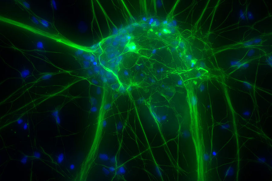
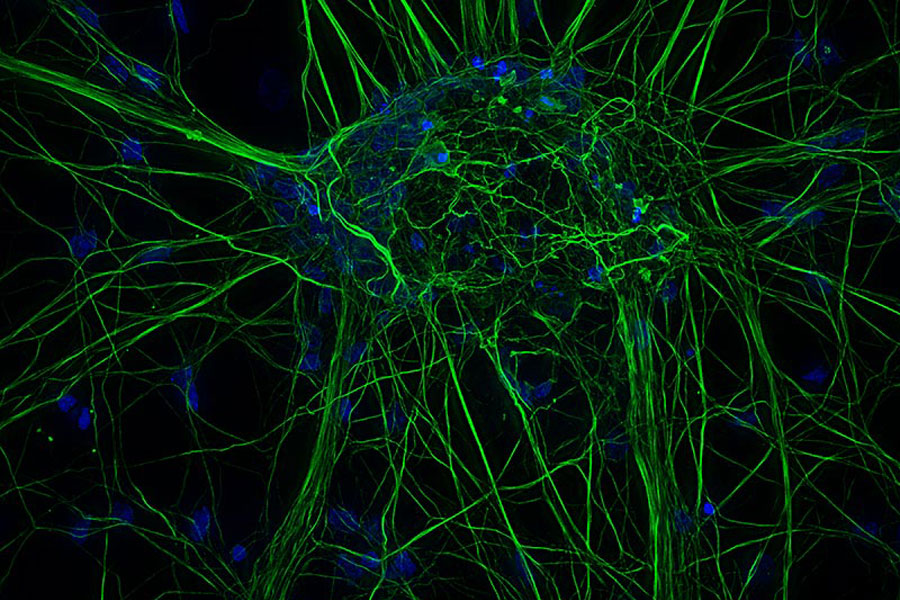
Widefield THUNDER Imager

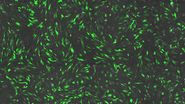
배양된 피질 뉴런입니다. 평면 59개의 Z-stack(두께: 21µm). 샘플 제공: FAN GmbH, Magdeburg, Germany.

관련 문서

Microscopy for neuroscience research

What are the Challenges in Neuroscience Microscopy?

eBook outlining the visualization of the nervous system using different types of microscopy techniques and methods to address questions in neuroscience.
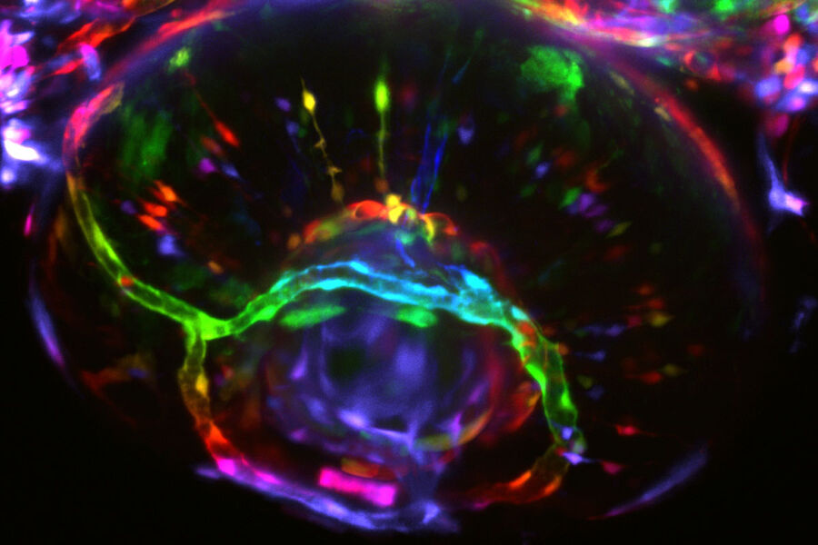
Virally labeled neurons (red) and astrocytes (green) in a cortical spheroid derived from human induced pluripotent stem cells. THUNDER Model Organism Imagerwith a 2x 0.15 NA objective at 3.4x zoomwas used to produce this 425 μm Z-stack (26 positions), which is presented here as an Extended Depth of Field(EDoF)projection.

Neuroscience Images

Neuroscience commonly uses microscopy to study the nervous system’s function and understand neurodegenerative diseases.
The role of extracellular signalling mechanisms in the correct development of the human brain

How do Cells Talk to Each Other During Neurodevelopment?

Professor Silvia Capello presents her group’s research on cellular crosstalk in neurodevelopmental disorders, using models such as cerebral organoids and assembloids.
Brain organoid section (DAPI) acquired using THUNDER Imager Live Cell. Image courtesy of Janina Kaspar and Irene Santisteban, Schäfer Lab, TUM.

Imaging Organoid Models to Investigate Brain Health

Imaging human brain organoid models to study the phenotypes of specialized brain cells called microglia, and the potential applications of these organoid models in health and disease.

신경과학 연구를 위한 현미경 방식

신경계에 대한 연구는 일반적으로 이벤트 및 구조의 고해상도 이미징을 위해 공초점 현미경에 의존합니다. 더 깊은 체내 이미징을 위해서는 다광자 현미경을 사용합니다. 근적외선 여기 광을 사용하는 기능으로 빛의 산란을 감소시켜, 최소의 침입성으로 깊은 이미징을 가능하게 하기 때문입니다. LightSheet 현미경 또한 광민감 샘플이나 3D 샘플에 선호됩니다. 이는 고유한 광학 단면 절편과 3D 이미징을 제공하면서 광독성을 줄입니다.

  • 광유전학은 빛을 이용해 신경 활동을 제어하는 기술을 말하며 이를 사용해 특정 신경망과 세포 신호를 연구할 수 있습니다. 이 방식에는 신경 세포막에 있는 광민감 단백질의 발현이 필요합니다. 광유전학을 밀리세컨드의 정밀 유리화와 결합하여 나노 단위로 이벤트를 탐색하는 것은 동적 과정 내에서 특정 시점을 연구할 수 있는 유망한 기술입니다.
  • 전기생리학은 조직과 세포의 전기적 특성에 대한 연구로, 뉴런의 전기적 특성에 대한 연구를 포함합니다. 신경과 근육 세포의 기능은 이온 채널을 통해 흐르는 이온 전류에 의존합니다. 이온 채널을 조사하는 한 가지 방법은 패치 클램핑을 사용하는 것입니다. 이 방법을 사용하면 이온 채널을 자세히 조사하고 뉴런처럼 주로 흥분할 수 있는 세포들을 비롯한 다양한 종류의 세포들의 전기 활동을 기록할 수 있습니다.

관련 문서

How did Laser Microdissection enable Pioneering Neuroscience Research?

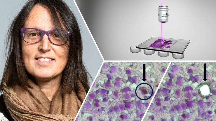
Dr. Marta Paterlini, a Senior Scientist at the Karolinska Institute, shares her experience of using laser microdissection (LMD) in groundbreaking research into adult human neurogenesis and offers…
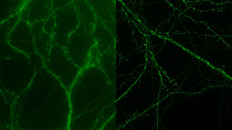
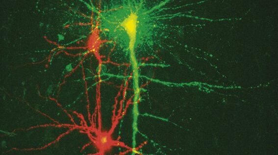
Mouse cortical neurons. Transgenic GFP (green). Image courtesy of Prof. Hui Guo, School of Life Sciences, Central South University, China

How Microscopy Helps the Study of Mechanoceptive and Synaptic Pathways

In this podcast, Dr Langenhan explains how microscopy helps his team to study mechanoceptive and synaptic pathways, their challenges, and how they overcome them.

Bridging Structure and Dynamics at the Nanoscale through Optogenetics and Electrical Stimulation

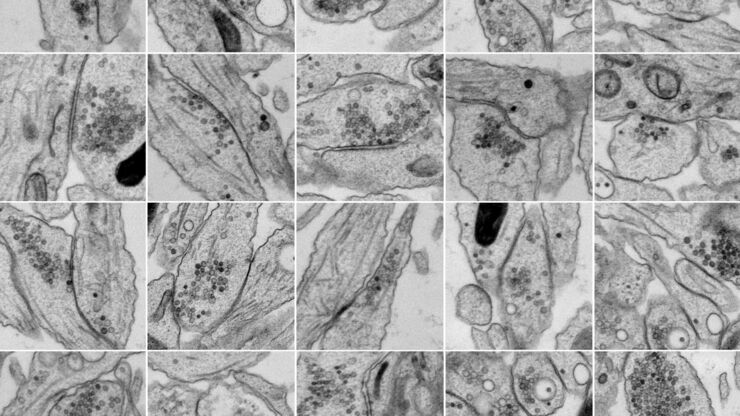
Nanoscale ultrastructural information is typically obtained by means of static imaging of a fixed and processed specimen. However, this is only a snapshot of one moment within a dynamic system in…
Image of murine dopaminergic neurons which have been marked for laser microdissection (LMD).

Neuron Isolation in Spatial Context with Laser Microdissection (LMD)

After Alzheimer’s disease, Parkinson’s is the second most common progressive neurodegenerative disease. Before the first symptoms manifest, up to 70% of dopamine-releasing neurons in the mid-brain…

New Standard in Electrophysiology and Deep Tissue Imaging

The function of nerve and muscle cells relies on ionic currents flowing through ion channels. These ion channels play a major role in cell physiology. One way to investigate ion channels is to use…
Scroll to top