HCS LSI Zebrafish High Content Screening Automation
Zebrafish
Leica HCS LSI enables new perspectives of whole animal research. Large filed of view, extended working distance and adaptive zoom optics provide new insights into large specimen.
Danio rerio. Blood vessels in the fin at high resolution (right) and zoom in on detailed Zebrafish eye.
Courtesy of D.…
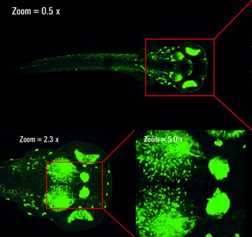
Zebrafish, Brain (in vivo)
Transgenic embryo, Danio rerio; GFP; Epithalamus; Optic Tectum. The images illustrate the zoom flexibility of Leica HCS LSI: The optical zoom is used first, then a combination of optical and confocal zoom is applied. The image shows a maximum projection of a 3D stack using…
Advanced Time Lapse: 4D Single Well Experiment Zebrafish Development
Tracking the development of life over time offers exciting insights for embryogenesis – all in the same specimen. From egg to embryo, obtain exciting views of organ development with Leica HCS LSI software and see the backbone formation during the growth of a zebrafish.
Zebrafish, novocord…
High content screening in 4D
Multiwell assay
Brain research: automated 4D imaging discloses the spatio-temporal dynamics of fluorescent signals throughout development.
4D series of 2 day old zebrafish embryos, Danio rerio. Ventral views of embryonic brains of the ETvmat2:gfp stable transgenic line were automatically acquired…


