TCS MP5 Optimized for Multiphoton Imaging
Deep Tissue Imaging
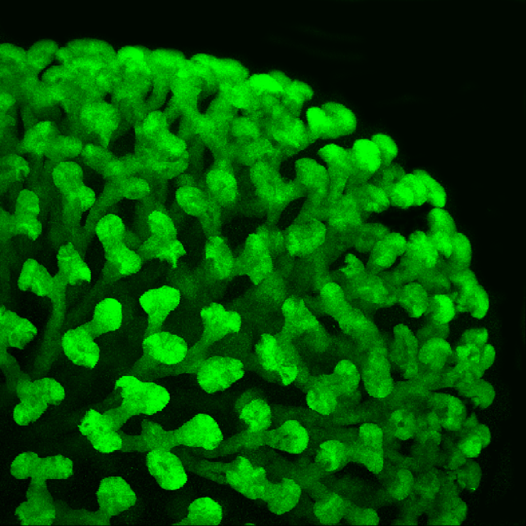
Ureteric bud from an embryonic day 16 kidney from a HoxB7 EGFP mouse.
Courtesy of Prof. Deborah Hyink, Mount Sinai School of Medicine, New York, NY, USA
Simultanous excitation with OPO and Ti:Sa
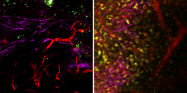
Mouse mammary gland (left) and spleen (right). Blood vessels labelled with 70kD-Texas Red excited with OPO at 1150 nm (red). Simultaneous excitation at 800 nm results in second harmonic generation (SHG) signal of type I collagen (purple) and autofluorescence of single cells (green).
Courtesy of…
Rapid acquisition of z-stacks in living animals
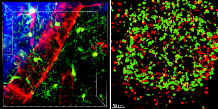
3D reconstructions of representative 50 µm z-stacks from timelapse acquisitions. Excitation at 910 nm, spectral unmixing was performed using the LAS AF software.
Left: Microglia labelled with GFP (green) are shown in relation to blood vessels injected with 655 nm quantum dots (red) residing in the…
Ultarapid imaging of embryonic blood flows
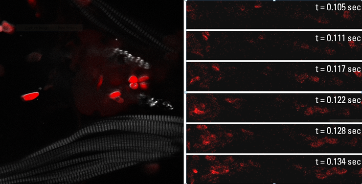
Zebrafish embryonic heart,100 µm deep. Blood cells labelled with DsRed (red), SHG of muscle (gray).
blood cells labelled with DsRed. The timelapse on the right was acquired with the resonant scanner at 167 frames/second at 512 x 64 pixels. Multiphoton excitation at 1100 nm with OPO.
Courtesy of Dr.…


