STELLARIS CRS Coherent Raman Scattering Microscope
Explore label-free chemical microscopy
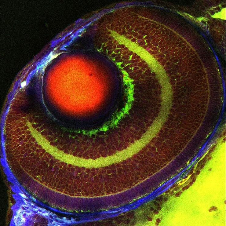
Label-free Vibrational Imaging of Cellular Structures in an Intact Zebrafish
This overlay image shows the eye of an intact zebrafish.
Green: Stimulated Raman scattering (SRS) image of Lipid components (at 2850 cm-1). Red: SRS image of Protein components (at 2935 cm-1). Blue: second-harmonic signals, mainly from the sclera and cornea.
The different layers of the retina…
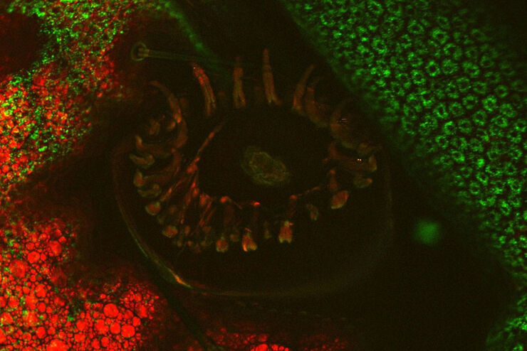
Biology – in vivo imaging of food moth (Plodia interpunctella)
This overlay image shows the surface of a larval silk gland.
Red: Lipid components (at 2850 cm-1). Green: autofluorescence of chitin at 1064 nm (wavelength).
The middle top of the image shows a bristle of the larvae.
Objective: HCX IR APO L25x/0.95 W 0.17.
Label-free Imaging of Mouse Brain Tissue Exhibiting Alzheimer’s Pathology
This movie shows a z-stack of a 50-μm thick mouse brain slice.
Green: CARS image of Lipid components (at 2850 cm-1) Blue: autofluorescence of intracellular vesicles likely containing lipofuscin
Myelinated neurites (lipid-rich tubular structures) and pathological lipid deposits (blob-shaped…


