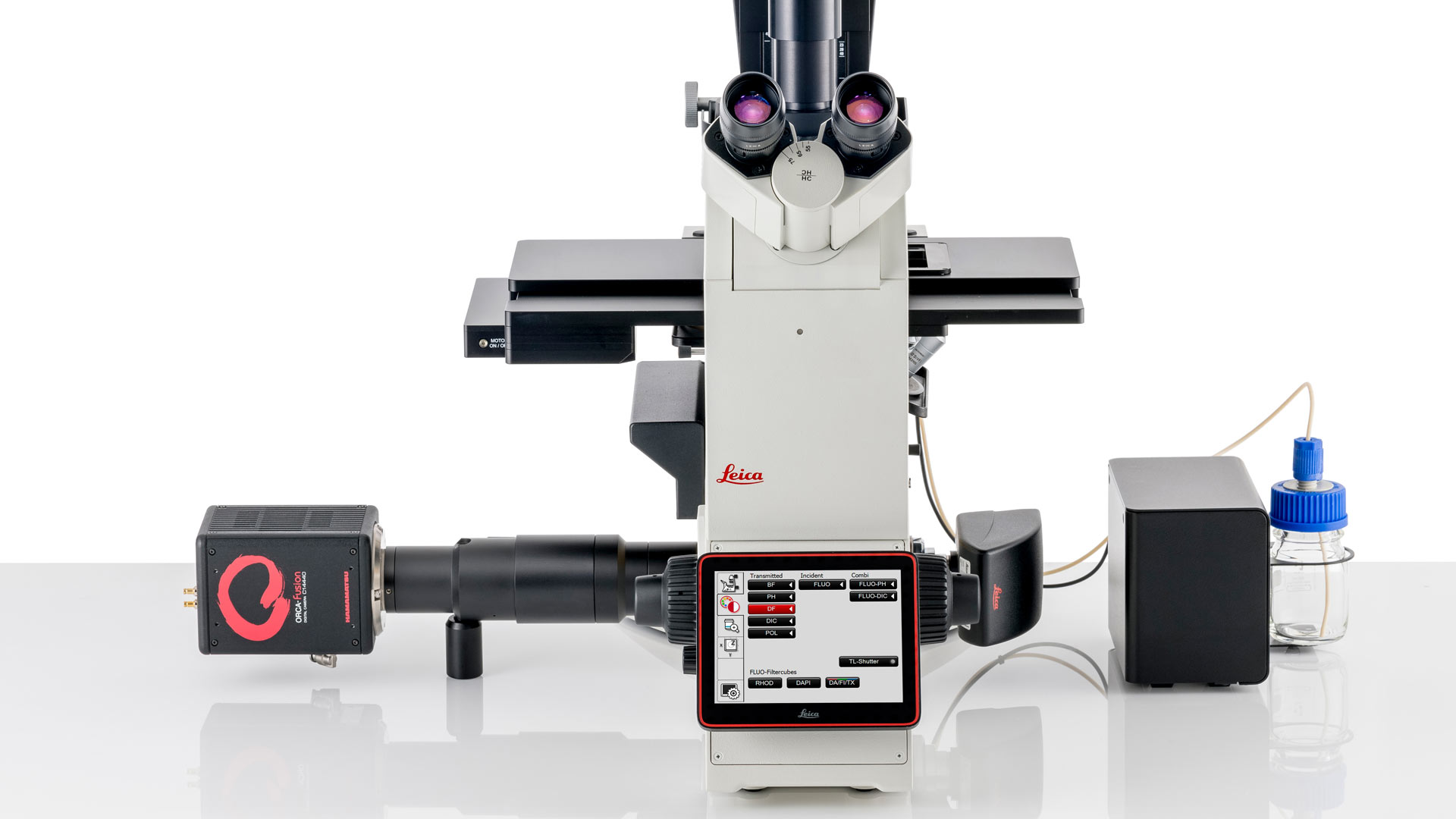
DMi8 Inverted Microscope
Streamline your complex microscopy workflows with the DMi8 inverted microscope. The platform enables you to reliably generate high quality data with solutions that you can tailor to your research requirements and budget.
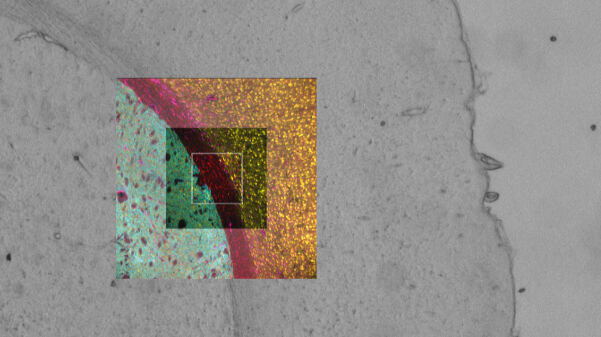
Enhanced Navigator integration
Easily switch from sample overview images to multi-channel view mode, giving your data spatial context throughout your workflow.
With enhanced Navigator integration, you can set up and perform complex multi-position experiments on a wide range of sample formats using templates for slides, dishes and multi-well plates.
Navigator combined with Adaptive Immersion technology allows you to seamlessly switch from overview images to high-resolution images using immersion objectives, without removing the sample from the stage.
Adaptive immersion technology
Patented and reliable Adaptive Immersion technology:
- Employs an embedded sensor to monitor and maintain immersion throughout your experiments.
- Enables you to use optimum water immersion objectives for live cell experiments under physiologically relevant conditions.
- Creates smoother workflows through hassle-free switching to immersion objectives.
Confidently gain more insights
Experience smoother and more efficient workflows with the DMi8
- Seamless integration of hardware triggering for maximum efficiency
- Faster scanning over large areas by combining 22 mm FOV optics with advanced cameras and the Quantum Stage
Convincing data from 3D samples
Generate better quality data every time regardless of your experience level. Extract the most insights from your 3D samples with our advanced technology suite including
- THUNDER
- SmartCORR
- Spinning Disk Confocal Scanner
- TIRF


Acquire precise and reproducible live cell data
Extract better data from your sample within seconds with SmartCORR, which optimizes your objective settings. This gives you the best possible data every time, with minimal effort.

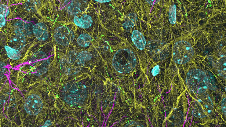
Beyond deconvolution - See through the haze
Elevate your DMi8 image quality with THUNDER, an opto-digital technology that uses the Computational Clearing method to generate high resolution, high contrast images, by removing the out of focus blur inherent to widefield images.
A simple yet powerful real time image enhancement tool, you can use THUNDER on both widefield and spinning disk data sets and see stunning results. With THUNDER Live you can optimize parameters with immediate visual feedback, improving data quality and reducing time to optimal results.
 biology.")
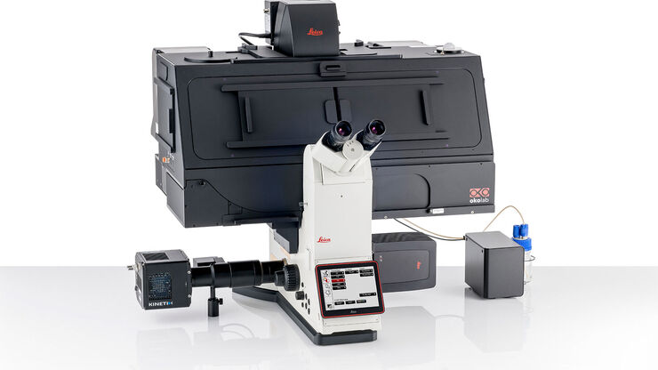
Customizable and upgradable
Future-proof your experiments with this adaptable solution. The DMi8's modular structure allows you to tailor your system to fit your current needs and budget.
As your research evolves, upgrade your system by adding features such as
- THUNDER
- Adaptive Focus Control (AFC)
- Climate control
- TIRF
- Spinning disk modules
Get the most from your DMi8
Maximize your investment through partnership with our Application Support Specialists throughout your system's lifetime.
Whether you are trying to get started or need to further tweak your system to address new workflows, our expert team will ensure you get the best possible data out of your system.

Contextualize your data to see the full picture
Efficiently set up your experiments with the DMi8’s intelligent automation, allowing you to focus more on your research questions. Gain relevant insights with:
- Automated Sample Finder - Quickly gain a comprehensive view of your sample putting your data into context.
- Fully Integrated Navigator Experience - Move through your experiments with ease.
- Adaptive Immersion technology – Confidently use water immersion objectives for the most challenging live cell experiments.
- 22 mm Field of View (FOV) – The throughput you need for enhanced data collection.

Automated Sample Finder
Simply place your sample on the stage and generate an in-focus overview of the entire sample at the click of a button with Automated Sample Finder.
- Eliminate the time and frustration of searching for a focused image of your sample, reducing your time to result.
- Gain context by understanding where specific data sets have been acquired from within your sample.
