Leica DM3000 & DM3000 LED
Upright Microscopes
Light Microscopes
Products
Home
Leica Microsystems
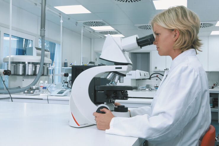
Leica DM3000 & DM3000 LED Uniquely Ergonomic System Microscopes With Intelligent Automation
Enhance Laboratory Workflow
Read our latest articles
A Guide to Differential Interference Contrast (DIC)
A DIC microscope is a widefield microscopy which has a polarization filter and Wollaston prism between the light source and condenser lens and also between the objective lens and camera sensor or…
A Guide to Phase Contrast
A phase contrast light microscope offers a way to view the structures of many types of biological specimens in greater contrast without the need of stains.
A Guide to Darkfield Microscopes
A darkfield microscope offers a way to view the structures of many types of biological specimens in greater contrast without the need of stains.
Differential Interference Contrast (DIC) Microscopy
This article demonstrates how differential interference contrast (DIC) can be actually better than brightfield illumination when using microscopy to image unstained biological specimens.
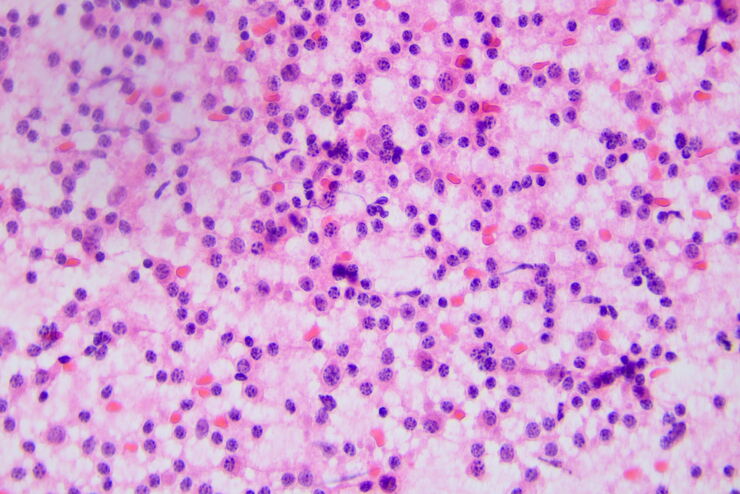
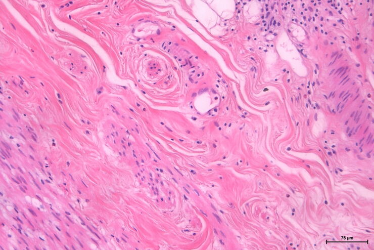
H&E Staining in Microscopy
If we consider the role of microscopy in pathologists’ daily routines, we often think of the diagnosis. While microscopes indeed play a crucial role at this stage of the pathology lab workflow, they…
How to Benefit from Digital Cytopathology
If you have thought of digital cytopathology as characterized by the digitization of glass slides, this webinar with Dr. Alessandro Caputo from the University Hospital of Salerno, Italy will broaden…
Factors to Consider when Selecting Clinical Microscopes
What matters if you would like to purchase a clinical microscope? Learn how to arrive at the best buying decision from our Science Lab Article.
The Time to Diagnosis is Crucial in Clinical Pathology
Abnormalities in tissues and fluids - that’s what pathologists are looking for when they examine specimens under the microscope. What they see and deduce from their findings is highly influential, as…
Perform Microscopy Analysis for Pathology Ergonomically and Efficiently
The main performance features of a microscope which are critical for rapid, ergonomic, and precise microscopic analysis of pathology specimens are described in this article. Microscopic analysis of…
Koehler Illumination: A Brief History and a Practical Set Up in Five Easy Steps
In this article, we will look at the history of the technique of Koehler Illumination in addition to how to adjust the components in five easy steps.
Fields of Application
Clinical Pathology
Discover how Leica pathology microscope solutions help clinical pathologists diagnose infections and diseases from bodily fluids and tissues.
Microscopy in Pathology
Analysis of specimens for pathology sometimes requires long hours working with a microscope. The result for the user may be physical discomfort and strain that can lead to reduced efficiency and the…
Anatomic Pathology
Learn how anatomic pathology microscopes from Leica Microsystems support efficient and accurate medical diagnoses.
