
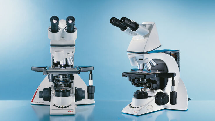
Leica DM3000 & DM3000 LED Uniquely Ergonomic System Microscopes With Intelligent Automation
The Leica DM3000 microscopes for pathology, cytology, haematology and many other applications feature a motorized nosepiece, condenser head, automated light intensity adjustment, and optional foot pedal. These intuitive microscopes improve workflows significantly. For special diagnostics requirements, the microscope is certified for in-vitro-diagnostics (IVD) like in-vitro-fertilization (IVF).
Intelligent and Innovative – The automated Leica DM3000 & DM3000 LED
Even faster, more comfortable, and more efficient: the Leica DM3000 microscopes combine operating convenience with an ergonomic design. The automated Leica DM3000 & DM3000 LED optimize work processes while adapting to each user’s physical requirements.
- Quickly change specimens with Leica Microsystems’ slide holders, designed so that slides can be changed in a single motion with one hand.
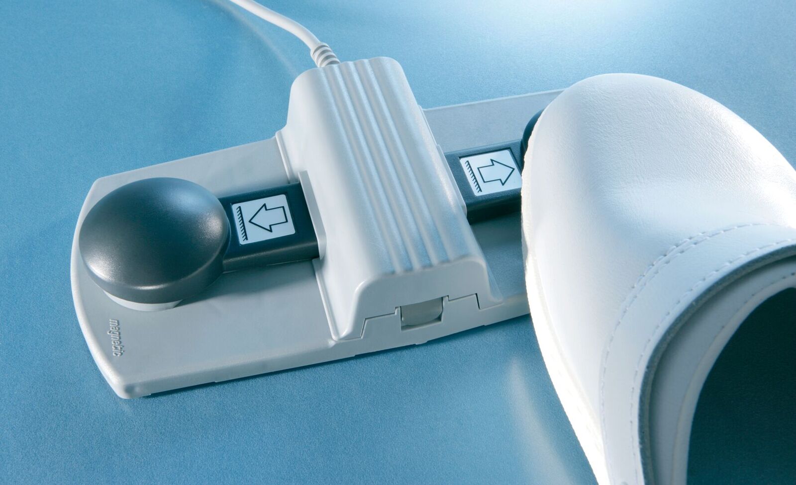
- Focus and stage adjustments can be performed with just one hand. The other hand remains free for other activities such as operating the PC.
- The modern stage design is rounded with no protruding parts. The entire design is compact and requires minimal space.
- The stage is built to last with a hard ceramic surface that is durable enough to take years of demanding use.
- The LED transmitted light illumination provides constant color temperature at all light intensity levels, clear differentiation of colors and is easy on the eyes – for reliable results and less fatiguing work. Due to the low power consumption of LED, the system is energy efficient.


Faster, Easier, More Efficient and Greater Comfort
Many laboratory activities promote poor posture, which can result in muscular tension and pain. But when it comes to microscopy, this discomfort is history: the Leica DM1000 – 3000 microscopes adjust perfectly to the physical needs of individual users – quickly, easily, and with minimal effort. They are designed to help prevent poor posture and so that the user can maintain a high level of concentration for long work sessions. Comfortable microscope use also promotes higher productivity in the laboratory.
Concentration Depends on Comfort
- Unique right-left handed operation* allows the controls to be switched over from the right- to the left-hand side of the microscope quickly and easily. Only one hand is required for focusing, and a user can decide which hand to leave free for other activities such as taking notes
- The user’s neck remains relaxed while viewing through the eyepieces. A choice of flexible adjustment or a fixed viewing angle of 15°, various tube lengths and convenient height adjustment accessories adapt the microscope to the individual user.
- Users automatically adopt a natural, comfortable position, even after extended periods of work. The symmetrical arrangement of the stage and focus controls helps to promote the users comfort at the microscope.
- The patented height adjustable focus knobs** can match the size of the user’s hands for a relaxed hand and arm position – a unique advance in microscope design. The adjustment can be made in seconds, which eliminates the need for wrist supports.
- The user’s seat height can be accommodated with the optional ErgoLift or ergomodules. Both options represent a small investment for a major gain in comfort.
* Patented DE 10 2004 053 437 B4; US 7,283,295; JP 4886995; US 7,330,306; CN 100445795
** Patented DE 103 40 721 B3; CN 100538430 C; JP 4677213 B2; US 7,233,435
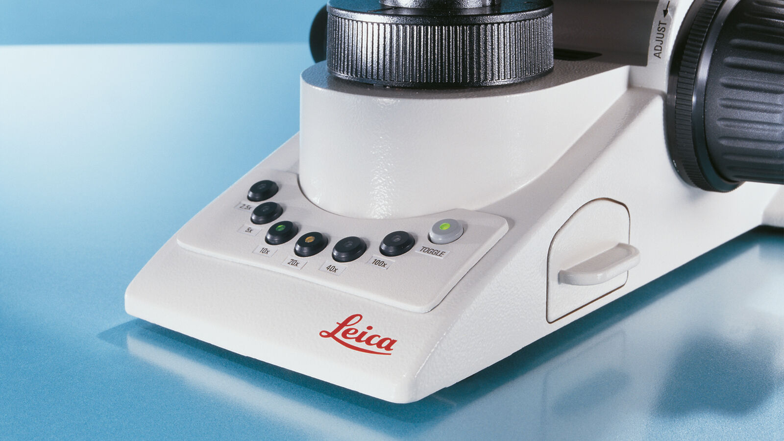
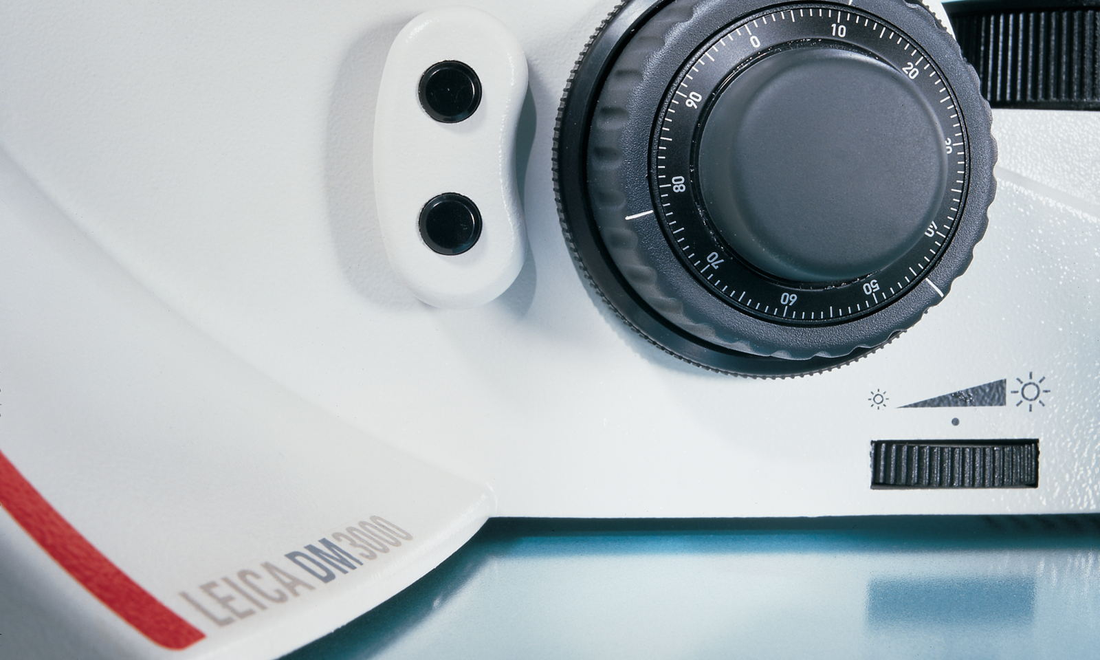
Change Objectives at the Touch of a Button
Leica’s motorized objective turret allows to change magnification in only half a second. Two buttons, conveniently located behind the focus knobs, or six buttons on the base of the microscope control the turret. Leica’s perfect toggle mode: any two of the six objectives can be assigned to the buttons behind the focus knobs. The remaining four objectives are available at the touch of a button whenever a different magnification is required. Six buttons on the base of the microscope are assigned to the six objectives. An optional foot pedal is also available, which frees the user’s hands for activities such as taking notes.
*Patented DE 10 2005 013 152 B4; US 7,570,421
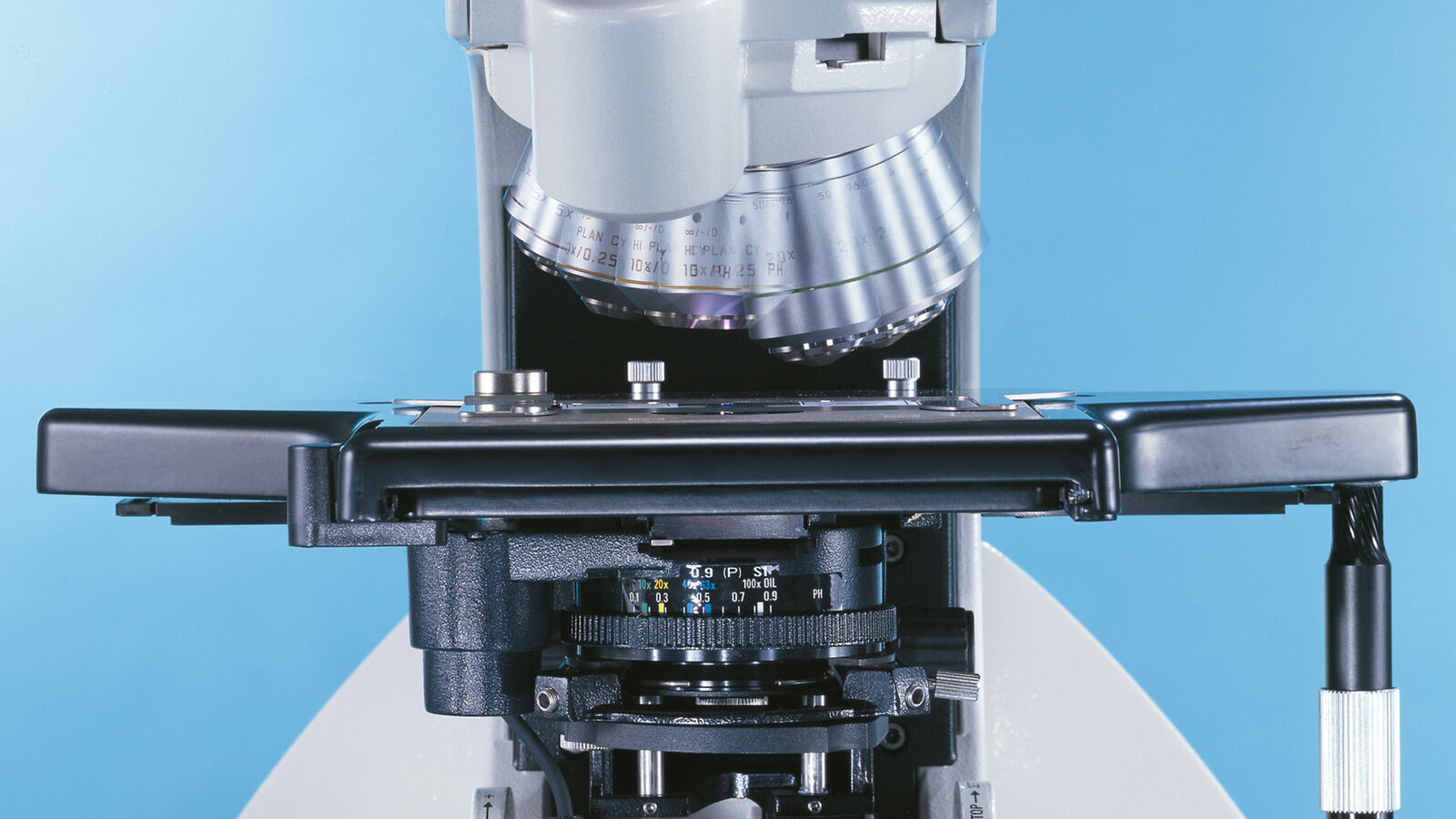
Automated Condenser Head
The automated condenser head automatically swings out when using objectives with less than 10x magnification and swings back into position when a higher magnification is selected. The microscope knows the best condenser position for every objective. For special applications, the user can individually adjust the condenser position by assigning this position to a control button. Leica’s automated condenser head accelerates workflow in the laboratory.
Microscopy in a New Light
The Leica DM1000 LED, DM2000 LED, DM2500 LED and DM3000 LED microscopes offer additional convenience with long-life LED transmitted light illumination. LED illumination provides constant color temperature at all light intensity levels without heating up the specimen. The LED’s high light density and optimal color reproduction provides brilliant images with a clear differentiation of the colors in the sample. The Leica DM2500 LED with LED lamphousing garantees ultra-bright LED illumination, ideal for highly light-absorbing specimens and contrasting methods like DIC. For the Leica DM1000 LED, Leica offers an optional portable version for field-based use.
Bulb Replacement is History: With extremely long lifetime LED of at least 50.000 hours, the LED illumination is very cost-effective, as frequent bulb-exchanges are no longer necessary.
Automatic Light Intensity Adjustment
The Leica DM3000 and DM3000 LED automatically set the light intensity to the appropriate level for any given magnification. The most recently used values are stored for each objective. The brightness impression remains constant for the observer and strong intensity changes are avoided for extended working sessions without eyestrain.

Brilliance Wherever You Look
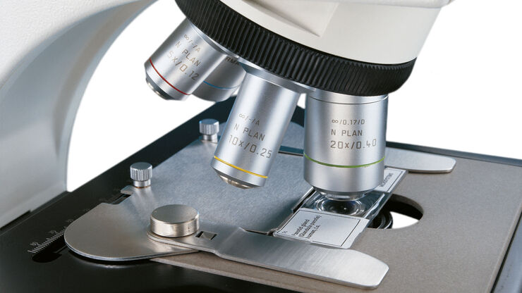
The Optical Features You Need
Razor-sharp contrast, precise contours, and brilliant fluorescence that reveals even the finest structures of dimly illuminated specimens; in terms of optical brilliance, the Leica DM1000 – 3000 series leaves nothing to be desired. Leica Microsystems offers a wide range of objectives – from planachromat with the best possible field flattening to apochromat with the highest resolution – and the ability to use sophisticated contrast methods.
- HI PLAN Planachromat objective series delivers images of astonishing clarity with significantly improved image flattening and chromatic correction.
- HI PLAN SL planachromat objective series maintains the brightness level at 4x, 10x, and 40x magnifications and preserves your preferred color impression. Continual adjustment of illumination intensity is history with Leica Microsystems SL (Synchronized Light) objectives.
- Special HI PLAN CY 10x/0.25 objectives feature excellent field flattening and color correction, and offer a long working distance of 12mm. These objectives are also available in an SL (Synchronized Light) version.
- A quick overview can be obtained with all models using the optional 1.25x screening objective.
The choice is yours: Use objectives from any Leica Microsystems` performance class, including our high-performance objectives from PL Fluotar class to Leica Apochromats with PL APO class for superior imaging quality. The Leica DM3000 and DM3000 LED feature an automated six-position objective turret.

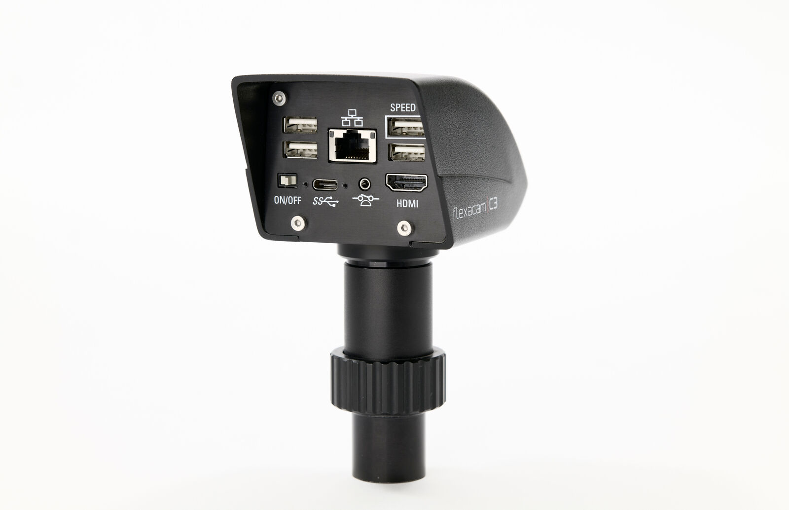
Leica Microsystems’ Premium Digital Cameras Open New Research Possibilities
Digital recording is beneficial for research. Digitized images can be analyzed for data not easily seen by the human eye. Leica Microsystems’ digital microscope cameras provide razor-sharp, brilliant images with uncompromising color fidelity. Leica Microsystems’ offers a wide variety of application-specific cameras to address your imaging requirements.
Full line of digital cameras features ease of use, image clarity, and excellent color fidelity – everything needed for precise image analysis, documentation, and reporting. For fluorescence photography, Leica Microsystems has developed digital cameras that deliver brilliant images from even very faint fluorescence specimens. High-end digital cameras capture the finest structures and most subtle color nuances and are suitable for all contrast methods, brightfield and darkfield, and even dimly illuminated specimens.
Leica DMshare V3
Make your Camera Wireless and Share Microscope Images on iPad and Android-Tablets. Leica DMshare provides a live display of what you and the microscope camera can see wirelessly on one or multiple tablets.
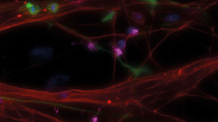

Fluorescence without compromise
The Leica DM1000 – 3000 microscopes are optionally available with a durable, high-quality fluorescence axis. The fluorescence axis for the Leica DM2000 – DM3000 features five filter cube positions on an easy-to-turn disk. If more filter cubes are required, the convenient quick-release allows replacement cubes to be snapped into place. The integrated neutral filter allows intensity reduction, which protects the specimen. The Leica DM1000 and DM1000 LED feature three filter cube positions on a slider.
Fluorescence filter cubes
The fluorescence axis of the Leica DM2000 – DM3000 is designed to accommodate all filter cubes of Leica Microsystems’ high-end research microscope range. The range of life science applications is covered completely, from routine FITC to GFP markers. The is no more need for additional BG38 filters because the new Leica K filter cubes integrate this capability. Leica Microsystems’ broad range of commonly used filter cubes is also available for the DM1000 and DM1000 LED.


