HCS A High Content Screening Automation
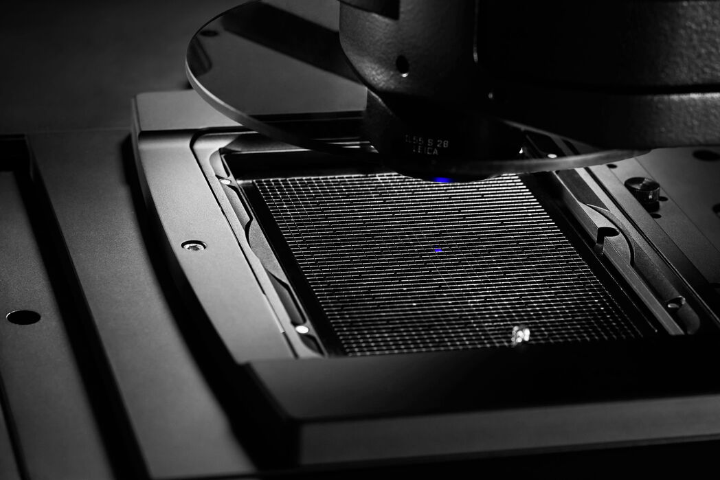
Breakthrough discoveries can happen when you are in the right place at the right time. Leica HCS A can speed up the process of discovery through high content screening automation, HCS A. The integration of Leica HCS A into the confocal and widefield systems of Leica Microsystems allows you to standardize biological applications for rapid and reproducible results.
Combine the flexibility of a point-scanning confocal with the high speed of a camera-based widefield system in the same assay to save time. Compensation for specimen drift, single object tracking and immersion fluid are taken care of for high quality raw data.
Leica HCS A supports microscope platforms of Leica Microsystems: Leica TCS SP8, Leica TCS SPE and LAS X Widefield Systems.
Regardless of which platform you choose Leica HCS A can mark the start of understanding of your biological question, too.

Key Features
Start thinking big
You can make strong inferences based on robust statistics by screening a large number of samples or conditions. Leica HCS A supports you with all the flexibility you need to incorporate any standard sample dish or multiwell plate. Since data is written to a storage device continuously image analysis can start immediately. Using OME-Tiff format maximizes compatibility and scalability. Tissues or organs typically won’t fit into one field of view. Leica HCS A offers a powerful stitching solution called Mosaic. You can combine these large images with time series, multiwell formats or custom positions. Leica HCS A will help you to tackle big questions while providing the full flexibility of a research microscope.
Start increasing your throughput
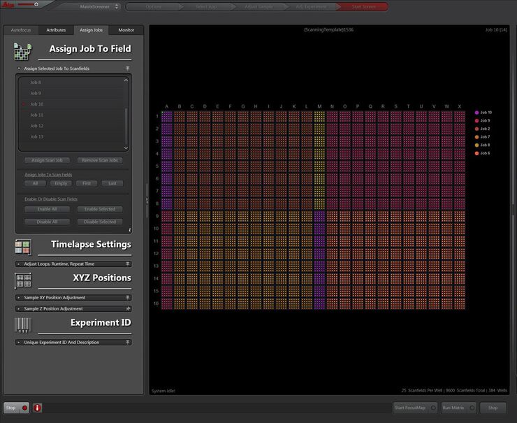
All image parameters as well as the geometry of the sample are adjusted in advance of the image acquisition by using scanning templates. These scanning templates decouple assay development from the high content screen to gain precious time. While the production environment continuous to churn out results new ideas can already be tested offline by the facility manager. Once established the imaging protocol becomes part of a scanning template ready for data recording. The scanning template documents all details of the acquisition process to be applied directly as well as for review by the researcher to ensure maximum data quality.
Start detecting rare events
The traditional paradigm of sifting through a large number of specimens manually cannot cope with rare events. Computer Aided Microscopy (CAM) allows these events to be continuously streamed to external storage devices using the OME-TIFF format where they are analysed in parallel to image acquisition. In conjunction with CAM, Leica HCS A can respond to feedback from analysis software about an event detected during acquisition. This powerful approach has proven to simplify large collaborative screening campaigns.


