Contact Us
Contact Us

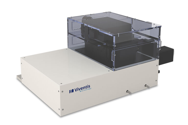
Viventis LS2 Live Dual View Light Sheet Fluorescence Microscope

Revealing life in its full context

Read our latest articles

Intestinal organoids label with FUCCI reporter to follow cell cycle dynamics. Courtesy of Franziska Moos. Liberali lab. FMI Basel (Switzerland).

Dual-View LightSheet Microscope for Large Multicellular Systems

Visualizing the dynamics of complex multicellular systems is a fundamental goal in biology. To address the challenges of live imaging over large spatiotemporal scales, Franziska Moos et. al. present…
Spheroid stained with Cyan: Dapi nuclear countertain; Green AF488 Involucrin; Orange AF55 Phalloidin Actin; Magenta AF647 CK14.

Notable AI-based Solutions for Phenotypic Drug Screening

Learn about notable optical microscope solutions for phenotypic drug screening using 3D-cell culture, both planning and execution, from this free, on-demand webinar.
Schematic graph of the light path in a Spalt-Ultramikroskop.

Confocal and Light Sheet Imaging

Optical imaging instrumentation can magnify tiny objects, zoom in on distant stars and reveal details that are invisible to the naked eye. But it notoriously suffers from an annoying problem: the…

Fields of Application

Live Cell Imaging

Shifting perspective from single microscope components to a full working live cell imaging solution, Leica Microsystems integrates microscope, LAS X imaging software, cameras, and dedicated…

Model Organisms in Research

A model organism is a species used by researchers to study specific biological processes. They have similar genetic characteristics to humans and are commonly used in research areas such as genetics,…

Organoids and 3D Cell Culture

One of the most exciting recent advancements in life science research is the development of 3D cell culture systems, such as organoids, spheroids, or organ-on-a-chip models. A 3D cell culture is an…
Scroll to top