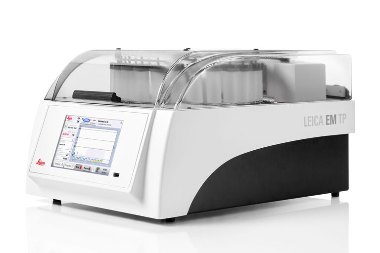
EM TP Automated Tissue Processor
Trust in the comparability of your results
Ensure the ultrastructures of your tissue samples can be precisely compared every time by preparing them with the EM TP tissue processor. With the EM TP you can be confident that tissue differences between samples observed with your light microscope (LM) or electron microscope (EM) are not caused by inconsistent manual preparation. The EM TP limits manual handling by at least 75% due to programmable, automatic processing of multiple samples.
Not only are samples in the same batch identically prepared, but the intuitive software also enables simple programming and reloading of entire protocols for reproducibility across batches. Additionally, the EM TP gives you the flexibility to stack various sizes of sample baskets so you can process different tissue types in one run.
Your benefits when working with the EM TP Tissue Processor:
- Eliminate inconsistencies that could be caused by manual sample preparation
- Ensure reproducibility across batches
- Process different tissue types in one run
- Maintain environmental conditions
Ensure comparability through automation
Eliminate the risk of inconsistencies that naturally occur when preparing your samples by hand. Thanks to the fully automated EM TP, manual handling is reduced by at least 75%. All processing steps of your experiment can be programmed with ease and are then executed by the system automatically. Trust in the comparability of your results and save valuable time with each run.
- Automatically distinguishes between EM or LM processing
- All experimental steps can be programmed, saved, and reloaded
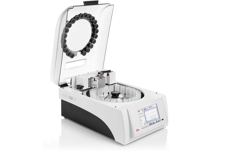
- Samples are transported automatically from vial to vial at the desired temperature and agitation speed
Reproducible experiments – reproducible results
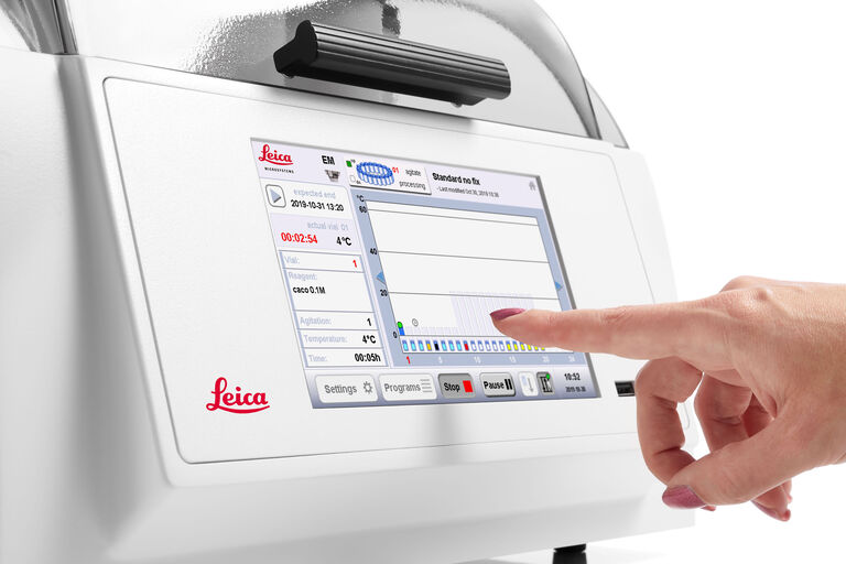
Ensure the reproducibility of your sample preparation within and in between runs as the sample preparation protocol can be programmed precisely. The integrated touch-screen-based software, guides you through the various setup options for simple programming of each step. After saving your programs, you can rest assured that your experiments are performed consistently every time.
With the EM TP you can program:
- All reagents that are used
- Duration of runs
- Environmental settings
- Delayed start and finish times
Process multiple tissues in one run
Compare multiple kinds of tissues with only one experimental setup.
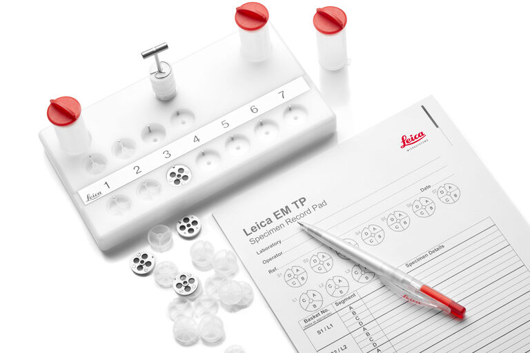
Various different-sized sample baskets give you the opportunity to flexibly stack your specimens and process them at the same time.
Increase your throughput and avoid wasting samples with every run.
Maintain environmental conditions
High quality processing is assured as the EM TP keeps your samples in a stable environment and controls the temperature precisely during preparation.
With the EM TP you can keep all reagents, which come in contact with the tissue, at the desired temperature. The heater and cooler system allows you to set and keep the temperature between +4 °C and +60 °C even during a delayed start or finish. For EM processing the system offers also pre-heat and pre-cool of the next reagent vial.
Choose your workflow according to your biological question
The EM TP tissue processor will help you get high quality results when conducting biological experiments ranging from standard TEM and SEM workflows to section tomography 3D workflows, to FIB SEM workflows, and many more.
For more information on workflow solutions for life science research, download our workflow booklet.
Standard SEM Workflow
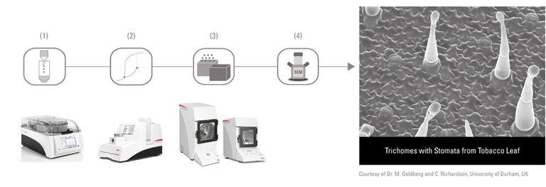
Investigate the surface architecture of chemically fixed samples with the standard SEM workflow. Prepare your samples with the EM TP tissue processer and critical point dry them afterwards with the EM CPD300. The next step is coating your samples with either the EM ACE200 or EM ACE600 followed by imaging in the SEM.
- Automated tissue processing (EM TP)
- Automated critical point drying (EM CPD300)
- Sputter and/or carbon coating (EM ACE200 / EM ACE600)
- Image analysis in the SEM
Section Tomography 3D Workflow
This workflow enables you to study the organization and interaction of biological structures within three dimensions in a defined volume. First process your samples at room temperature, followed by semi-thick serial sectioning on TEM grids. After the staining step, proceed with the image analysis in the TEM.
- Automated tissue processing (EM TP)
- Trimming (EM TRIM2)
- Serial sectioning (EM UC7)
- Staining (EM AC20)
- Image analysis in the TEM

